Arshia Yazdani, Mohammad Ranaee, Sara Babazadeh, Fatemeh Shafizadeh
{"title":"胎盘间充质发育不良伴严重宫内生长受限1例报告。","authors":"Arshia Yazdani, Mohammad Ranaee, Sara Babazadeh, Fatemeh Shafizadeh","doi":"10.30699/IJP.2023.548113.2828","DOIUrl":null,"url":null,"abstract":"<p><p>Placental mesenchymal dysplasia (PMD) is an uncommon placental lesion, which may mimic molar pregnancy at gross and microscopic examination. PMD can be associated with fetal growth restriction, Beckwith-Wiedemann syndrome, intrauterine fetal death, and preterm delivery. Nonetheless, it may also be associated with a normal appearing fetus. We aimed to emphasize that clinicians, radiologists, and pathologists should be aware of PMD as one of the etiologies of intrauterine growth restriction (IUGR). We presented the case of a 27-year-old gravida 1, para 1 woman who was admitted to Ayatollah Rouhani hospital, in Babol, Iran, at 30 weeks of gestation due to severe IUGR and fetal tachycardia. Ultrasound examination showed uteroplacental insufficiency and increased resistive index (RI) of umbilical artery. At last, a normal female fetus (1320 g) with no definitive anomalies was delivered by cesarean section. Pathological examination revealed cystically dilated stem villi with peripherally located thick-walled muscular stem vessels, and also stromal fibroblasts overgrowth in some stem villi. None of the examined sections revealed trophoblastic proliferation or stromal trophoblastic inclusion. The findings confirmed the diagnosis of PMD. Careful radiological and pathological examination should be performed in the case of IUGR for ruling out the rare placental abnormalities, including PMD.</p>","PeriodicalId":38900,"journal":{"name":"Iranian Journal of Pathology","volume":"18 2","pages":"221-224"},"PeriodicalIF":0.0000,"publicationDate":"2023-01-01","publicationTypes":"Journal Article","fieldsOfStudy":null,"isOpenAccess":false,"openAccessPdf":"https://www.ncbi.nlm.nih.gov/pmc/articles/PMC10439749/pdf/","citationCount":"0","resultStr":"{\"title\":\"Placental Mesenchymal Dysplasia Associated with Severe Intrauterine Growth Restriction: A Case Report.\",\"authors\":\"Arshia Yazdani, Mohammad Ranaee, Sara Babazadeh, Fatemeh Shafizadeh\",\"doi\":\"10.30699/IJP.2023.548113.2828\",\"DOIUrl\":null,\"url\":null,\"abstract\":\"<p><p>Placental mesenchymal dysplasia (PMD) is an uncommon placental lesion, which may mimic molar pregnancy at gross and microscopic examination. PMD can be associated with fetal growth restriction, Beckwith-Wiedemann syndrome, intrauterine fetal death, and preterm delivery. Nonetheless, it may also be associated with a normal appearing fetus. We aimed to emphasize that clinicians, radiologists, and pathologists should be aware of PMD as one of the etiologies of intrauterine growth restriction (IUGR). We presented the case of a 27-year-old gravida 1, para 1 woman who was admitted to Ayatollah Rouhani hospital, in Babol, Iran, at 30 weeks of gestation due to severe IUGR and fetal tachycardia. Ultrasound examination showed uteroplacental insufficiency and increased resistive index (RI) of umbilical artery. At last, a normal female fetus (1320 g) with no definitive anomalies was delivered by cesarean section. Pathological examination revealed cystically dilated stem villi with peripherally located thick-walled muscular stem vessels, and also stromal fibroblasts overgrowth in some stem villi. None of the examined sections revealed trophoblastic proliferation or stromal trophoblastic inclusion. The findings confirmed the diagnosis of PMD. Careful radiological and pathological examination should be performed in the case of IUGR for ruling out the rare placental abnormalities, including PMD.</p>\",\"PeriodicalId\":38900,\"journal\":{\"name\":\"Iranian Journal of Pathology\",\"volume\":\"18 2\",\"pages\":\"221-224\"},\"PeriodicalIF\":0.0000,\"publicationDate\":\"2023-01-01\",\"publicationTypes\":\"Journal Article\",\"fieldsOfStudy\":null,\"isOpenAccess\":false,\"openAccessPdf\":\"https://www.ncbi.nlm.nih.gov/pmc/articles/PMC10439749/pdf/\",\"citationCount\":\"0\",\"resultStr\":null,\"platform\":\"Semanticscholar\",\"paperid\":null,\"PeriodicalName\":\"Iranian Journal of Pathology\",\"FirstCategoryId\":\"1085\",\"ListUrlMain\":\"https://doi.org/10.30699/IJP.2023.548113.2828\",\"RegionNum\":0,\"RegionCategory\":null,\"ArticlePicture\":[],\"TitleCN\":null,\"AbstractTextCN\":null,\"PMCID\":null,\"EPubDate\":\"\",\"PubModel\":\"\",\"JCR\":\"Q3\",\"JCRName\":\"Medicine\",\"Score\":null,\"Total\":0}","platform":"Semanticscholar","paperid":null,"PeriodicalName":"Iranian Journal of Pathology","FirstCategoryId":"1085","ListUrlMain":"https://doi.org/10.30699/IJP.2023.548113.2828","RegionNum":0,"RegionCategory":null,"ArticlePicture":[],"TitleCN":null,"AbstractTextCN":null,"PMCID":null,"EPubDate":"","PubModel":"","JCR":"Q3","JCRName":"Medicine","Score":null,"Total":0}
Placental Mesenchymal Dysplasia Associated with Severe Intrauterine Growth Restriction: A Case Report.
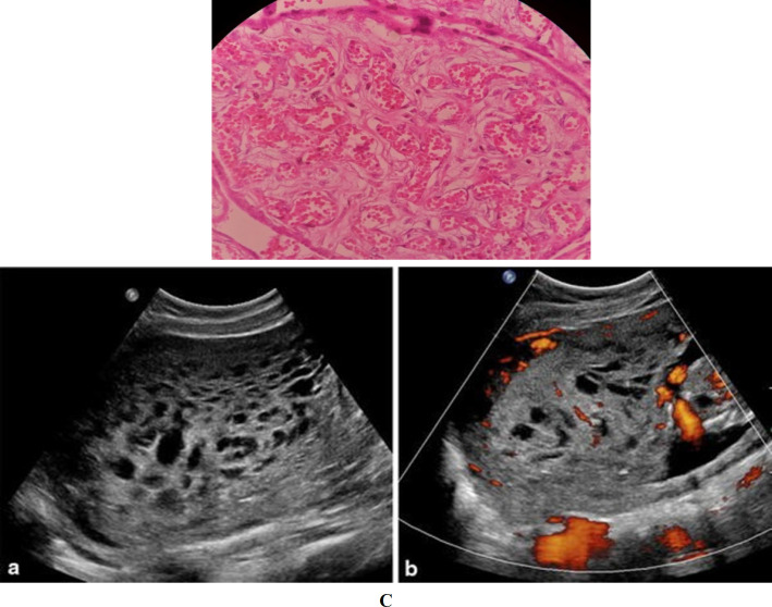
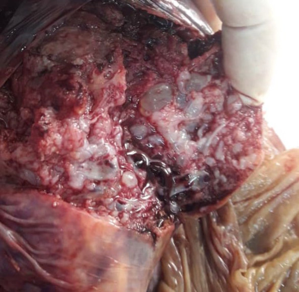
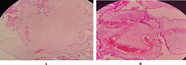
Placental mesenchymal dysplasia (PMD) is an uncommon placental lesion, which may mimic molar pregnancy at gross and microscopic examination. PMD can be associated with fetal growth restriction, Beckwith-Wiedemann syndrome, intrauterine fetal death, and preterm delivery. Nonetheless, it may also be associated with a normal appearing fetus. We aimed to emphasize that clinicians, radiologists, and pathologists should be aware of PMD as one of the etiologies of intrauterine growth restriction (IUGR). We presented the case of a 27-year-old gravida 1, para 1 woman who was admitted to Ayatollah Rouhani hospital, in Babol, Iran, at 30 weeks of gestation due to severe IUGR and fetal tachycardia. Ultrasound examination showed uteroplacental insufficiency and increased resistive index (RI) of umbilical artery. At last, a normal female fetus (1320 g) with no definitive anomalies was delivered by cesarean section. Pathological examination revealed cystically dilated stem villi with peripherally located thick-walled muscular stem vessels, and also stromal fibroblasts overgrowth in some stem villi. None of the examined sections revealed trophoblastic proliferation or stromal trophoblastic inclusion. The findings confirmed the diagnosis of PMD. Careful radiological and pathological examination should be performed in the case of IUGR for ruling out the rare placental abnormalities, including PMD.

 求助内容:
求助内容: 应助结果提醒方式:


