Daniel Strüder, Christoph Lachmann, Sara Maria van Bonn, Eberhard Grambow, Sebastian P Schraven, Robert Mlynski, Brigitte Vollmar
{"title":"背侧皮肤褶腔作为一种新的鼓膜伤口愈合模型:对上皮化伤口病理生理的活体观察。","authors":"Daniel Strüder, Christoph Lachmann, Sara Maria van Bonn, Eberhard Grambow, Sebastian P Schraven, Robert Mlynski, Brigitte Vollmar","doi":"10.1159/000519774","DOIUrl":null,"url":null,"abstract":"<p><strong>Background: </strong>Tympanic membrane perforations (TMPs) are a common complication of trauma and infection. Persisting perforations result from the unique location of the tympanic membrane. The wound is surrounded by air of the middle ear and the external auditory canal. The inadequate wound bed, growth factor, and blood supply lead to circular epithelialization of the perforation's edge and premature interruption of defect closure. Orthotopic animal models use mechanical or chemical tympanic membrane laceration to identify bioactive wound dressings and overcome premature epithelialization. However, all orthotopic models essentially lack repetitive visualization of the biomaterial-wound interface. Therefore, recent progress in 3D printing of customized wound dressings has not yet been transferred to the unique wound setup of the TMP. Here, we present a novel application for the mice dorsal skinfold chamber (DSC) with an epithelialized full-thickness defect as TMP model.</p><p><strong>Methods: </strong>A circular 2-mm defect was cut into the extended dorsal skinfold using a biopsy punch. The skinfold was either perforated through both skin layers without prior preparation or perforated on 1 side, following resection of the opposing skin layer. In both groups, the wound was sealed with a coverslip or left unclosed (n = 4). All animals were examined for epithelialization of the edge (histology), size of the perforation (planimetry), neovascularization (repetitive intravital fluorescence microscopy), and inflammation (immunohistology).</p><p><strong>Results: </strong>The edge of the perforation was overgrown by the cornified squamous epithelium in all pre-parations. Reduction in the perforation's size was enhanced by application of a coverslip. Microsurgical preparation before biopsy punch perforation and sealing with a coverslip enabled repetitive high-quality intravital fluorescence microscopy. However, spontaneous reduction of the perforation occurred frequently. Therefore, the direct biopsy punch perforation without microsurgical preparation was favorable: spontaneous reduction did not occur throughout 21 days. Moreover, the visualization of the neovascularization was sufficient in intravital microscopy.</p><p><strong>Conclusions: </strong>The DSC full-thickness defect is a valuable supplement to orthotopic TMP models. Repetitive intravital microscopy of the epithelialized edge enables investigation of the underlying pathophysiology during the transition from the inflammation to the proliferation phase of wound healing. Using established analysis procedures, the present model provides an effective platform for the screening of bioactive materials and transferring progress in tissue engineering to the special conditions of tympanic membrane wound healing.</p>","PeriodicalId":12222,"journal":{"name":"European Surgical Research","volume":"64 2","pages":"286-300"},"PeriodicalIF":1.9000,"publicationDate":"2023-01-01","publicationTypes":"Journal Article","fieldsOfStudy":null,"isOpenAccess":false,"openAccessPdf":"https://www.ncbi.nlm.nih.gov/pmc/articles/PMC9808650/pdf/","citationCount":"0","resultStr":"{\"title\":\"The Dorsal Skinfold Chamber as a New Tympanic Membrane Wound Healing Model: Intravital Insights into the Pathophysiology of Epithelialized Wounds.\",\"authors\":\"Daniel Strüder, Christoph Lachmann, Sara Maria van Bonn, Eberhard Grambow, Sebastian P Schraven, Robert Mlynski, Brigitte Vollmar\",\"doi\":\"10.1159/000519774\",\"DOIUrl\":null,\"url\":null,\"abstract\":\"<p><strong>Background: </strong>Tympanic membrane perforations (TMPs) are a common complication of trauma and infection. Persisting perforations result from the unique location of the tympanic membrane. The wound is surrounded by air of the middle ear and the external auditory canal. The inadequate wound bed, growth factor, and blood supply lead to circular epithelialization of the perforation's edge and premature interruption of defect closure. Orthotopic animal models use mechanical or chemical tympanic membrane laceration to identify bioactive wound dressings and overcome premature epithelialization. However, all orthotopic models essentially lack repetitive visualization of the biomaterial-wound interface. Therefore, recent progress in 3D printing of customized wound dressings has not yet been transferred to the unique wound setup of the TMP. Here, we present a novel application for the mice dorsal skinfold chamber (DSC) with an epithelialized full-thickness defect as TMP model.</p><p><strong>Methods: </strong>A circular 2-mm defect was cut into the extended dorsal skinfold using a biopsy punch. The skinfold was either perforated through both skin layers without prior preparation or perforated on 1 side, following resection of the opposing skin layer. In both groups, the wound was sealed with a coverslip or left unclosed (n = 4). All animals were examined for epithelialization of the edge (histology), size of the perforation (planimetry), neovascularization (repetitive intravital fluorescence microscopy), and inflammation (immunohistology).</p><p><strong>Results: </strong>The edge of the perforation was overgrown by the cornified squamous epithelium in all pre-parations. Reduction in the perforation's size was enhanced by application of a coverslip. Microsurgical preparation before biopsy punch perforation and sealing with a coverslip enabled repetitive high-quality intravital fluorescence microscopy. However, spontaneous reduction of the perforation occurred frequently. Therefore, the direct biopsy punch perforation without microsurgical preparation was favorable: spontaneous reduction did not occur throughout 21 days. Moreover, the visualization of the neovascularization was sufficient in intravital microscopy.</p><p><strong>Conclusions: </strong>The DSC full-thickness defect is a valuable supplement to orthotopic TMP models. Repetitive intravital microscopy of the epithelialized edge enables investigation of the underlying pathophysiology during the transition from the inflammation to the proliferation phase of wound healing. Using established analysis procedures, the present model provides an effective platform for the screening of bioactive materials and transferring progress in tissue engineering to the special conditions of tympanic membrane wound healing.</p>\",\"PeriodicalId\":12222,\"journal\":{\"name\":\"European Surgical Research\",\"volume\":\"64 2\",\"pages\":\"286-300\"},\"PeriodicalIF\":1.9000,\"publicationDate\":\"2023-01-01\",\"publicationTypes\":\"Journal Article\",\"fieldsOfStudy\":null,\"isOpenAccess\":false,\"openAccessPdf\":\"https://www.ncbi.nlm.nih.gov/pmc/articles/PMC9808650/pdf/\",\"citationCount\":\"0\",\"resultStr\":null,\"platform\":\"Semanticscholar\",\"paperid\":null,\"PeriodicalName\":\"European Surgical Research\",\"FirstCategoryId\":\"3\",\"ListUrlMain\":\"https://doi.org/10.1159/000519774\",\"RegionNum\":4,\"RegionCategory\":\"医学\",\"ArticlePicture\":[],\"TitleCN\":null,\"AbstractTextCN\":null,\"PMCID\":null,\"EPubDate\":\"\",\"PubModel\":\"\",\"JCR\":\"Q2\",\"JCRName\":\"SURGERY\",\"Score\":null,\"Total\":0}","platform":"Semanticscholar","paperid":null,"PeriodicalName":"European Surgical Research","FirstCategoryId":"3","ListUrlMain":"https://doi.org/10.1159/000519774","RegionNum":4,"RegionCategory":"医学","ArticlePicture":[],"TitleCN":null,"AbstractTextCN":null,"PMCID":null,"EPubDate":"","PubModel":"","JCR":"Q2","JCRName":"SURGERY","Score":null,"Total":0}
The Dorsal Skinfold Chamber as a New Tympanic Membrane Wound Healing Model: Intravital Insights into the Pathophysiology of Epithelialized Wounds.
Background: Tympanic membrane perforations (TMPs) are a common complication of trauma and infection. Persisting perforations result from the unique location of the tympanic membrane. The wound is surrounded by air of the middle ear and the external auditory canal. The inadequate wound bed, growth factor, and blood supply lead to circular epithelialization of the perforation's edge and premature interruption of defect closure. Orthotopic animal models use mechanical or chemical tympanic membrane laceration to identify bioactive wound dressings and overcome premature epithelialization. However, all orthotopic models essentially lack repetitive visualization of the biomaterial-wound interface. Therefore, recent progress in 3D printing of customized wound dressings has not yet been transferred to the unique wound setup of the TMP. Here, we present a novel application for the mice dorsal skinfold chamber (DSC) with an epithelialized full-thickness defect as TMP model.
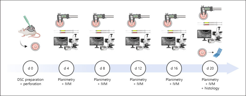
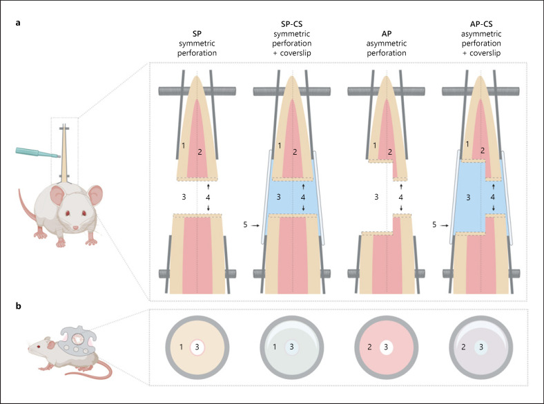
Methods: A circular 2-mm defect was cut into the extended dorsal skinfold using a biopsy punch. The skinfold was either perforated through both skin layers without prior preparation or perforated on 1 side, following resection of the opposing skin layer. In both groups, the wound was sealed with a coverslip or left unclosed (n = 4). All animals were examined for epithelialization of the edge (histology), size of the perforation (planimetry), neovascularization (repetitive intravital fluorescence microscopy), and inflammation (immunohistology).
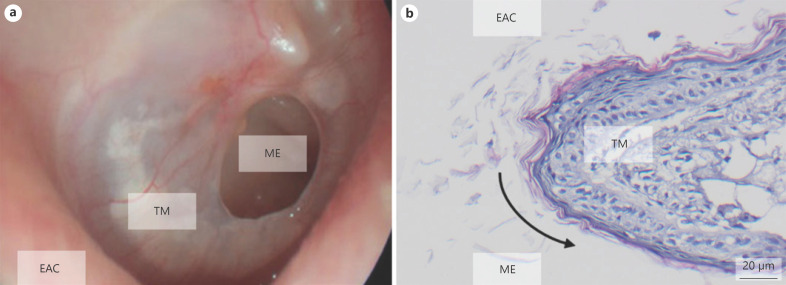
Results: The edge of the perforation was overgrown by the cornified squamous epithelium in all pre-parations. Reduction in the perforation's size was enhanced by application of a coverslip. Microsurgical preparation before biopsy punch perforation and sealing with a coverslip enabled repetitive high-quality intravital fluorescence microscopy. However, spontaneous reduction of the perforation occurred frequently. Therefore, the direct biopsy punch perforation without microsurgical preparation was favorable: spontaneous reduction did not occur throughout 21 days. Moreover, the visualization of the neovascularization was sufficient in intravital microscopy.
Conclusions: The DSC full-thickness defect is a valuable supplement to orthotopic TMP models. Repetitive intravital microscopy of the epithelialized edge enables investigation of the underlying pathophysiology during the transition from the inflammation to the proliferation phase of wound healing. Using established analysis procedures, the present model provides an effective platform for the screening of bioactive materials and transferring progress in tissue engineering to the special conditions of tympanic membrane wound healing.
期刊介绍:
''European Surgical Research'' features original clinical and experimental papers, condensed reviews of new knowledge relevant to surgical research, and short technical notes serving the information needs of investigators in various fields of operative medicine. Coverage includes surgery, surgical pathophysiology, drug usage, and new surgical techniques. Special consideration is given to information on the use of animal models, physiological and biological methods as well as biophysical measuring and recording systems. The journal is of particular value for workers interested in pathophysiologic concepts, new techniques and in how these can be introduced into clinical work or applied when critical decisions are made concerning the use of new procedures or drugs.
 求助内容:
求助内容: 应助结果提醒方式:


