Alberto Nieri, Luca Urso, Matteo Caracciolo, Maria Ciccone, Licia Uccelli, Corrado Cittanti, Antonio Cuneo, Mirco Bartolomei
{"title":"霍奇金淋巴瘤患者炎性FDG摄取的误读:1例报告。","authors":"Alberto Nieri, Luca Urso, Matteo Caracciolo, Maria Ciccone, Licia Uccelli, Corrado Cittanti, Antonio Cuneo, Mirco Bartolomei","doi":"10.22038/AOJNMB.2022.66011.1457","DOIUrl":null,"url":null,"abstract":"<p><p>Hodgkin Lymphoma (HL) is a malignancy involving lymph nodes and lymphatic system. [<sup>18</sup>F]F-FDG PET/CT (FDG-PET) imaging is routinely used for staging, to assess early chemotherapy response (interim FDG-PET), at the end of treatment (EoT FDG-PET) and for the identification of disease recurrence. We present a case of a 39-year-old man treated for HL. FDG-PET scans performed after first line therapy (both Interim PET and at the end of therapy) demonstrated a persistent and significant mediastinal FDG uptake. The patient was treated with a second line therapy but the FDG-PET uptake did not change. After board discussion a new surgical, thoracoscopy-guided biopsy was performed. Histopathology demonstrated a dense fibrous tissue with occasional chronic inflammatory infiltrates. Persistent FDG-PET positivity may suggest refractory or relapsed disease. However, occasionally, non-malignant conditions are responsible for a persistent FDG uptake, not related to primary disease. An accurate evaluation of clinical history and previous imaging exams is mandatory for clinicians and others experts to avoid misinterpretations of FDG-PET results. Nevertheless, in some cases, only a more invasive procedure, such as a biopsy, may finally lead to a definitive diagnosis.</p>","PeriodicalId":8503,"journal":{"name":"Asia Oceania Journal of Nuclear Medicine and Biology","volume":null,"pages":null},"PeriodicalIF":0.0000,"publicationDate":"2023-01-01","publicationTypes":"Journal Article","fieldsOfStudy":null,"isOpenAccess":false,"openAccessPdf":"https://www.ncbi.nlm.nih.gov/pmc/articles/PMC10261686/pdf/","citationCount":"0","resultStr":"{\"title\":\"Misinterpretation of an inflammatory FDG uptake in a patient treated for Hodgkin lymphoma: a case report.\",\"authors\":\"Alberto Nieri, Luca Urso, Matteo Caracciolo, Maria Ciccone, Licia Uccelli, Corrado Cittanti, Antonio Cuneo, Mirco Bartolomei\",\"doi\":\"10.22038/AOJNMB.2022.66011.1457\",\"DOIUrl\":null,\"url\":null,\"abstract\":\"<p><p>Hodgkin Lymphoma (HL) is a malignancy involving lymph nodes and lymphatic system. [<sup>18</sup>F]F-FDG PET/CT (FDG-PET) imaging is routinely used for staging, to assess early chemotherapy response (interim FDG-PET), at the end of treatment (EoT FDG-PET) and for the identification of disease recurrence. We present a case of a 39-year-old man treated for HL. FDG-PET scans performed after first line therapy (both Interim PET and at the end of therapy) demonstrated a persistent and significant mediastinal FDG uptake. The patient was treated with a second line therapy but the FDG-PET uptake did not change. After board discussion a new surgical, thoracoscopy-guided biopsy was performed. Histopathology demonstrated a dense fibrous tissue with occasional chronic inflammatory infiltrates. Persistent FDG-PET positivity may suggest refractory or relapsed disease. However, occasionally, non-malignant conditions are responsible for a persistent FDG uptake, not related to primary disease. An accurate evaluation of clinical history and previous imaging exams is mandatory for clinicians and others experts to avoid misinterpretations of FDG-PET results. Nevertheless, in some cases, only a more invasive procedure, such as a biopsy, may finally lead to a definitive diagnosis.</p>\",\"PeriodicalId\":8503,\"journal\":{\"name\":\"Asia Oceania Journal of Nuclear Medicine and Biology\",\"volume\":null,\"pages\":null},\"PeriodicalIF\":0.0000,\"publicationDate\":\"2023-01-01\",\"publicationTypes\":\"Journal Article\",\"fieldsOfStudy\":null,\"isOpenAccess\":false,\"openAccessPdf\":\"https://www.ncbi.nlm.nih.gov/pmc/articles/PMC10261686/pdf/\",\"citationCount\":\"0\",\"resultStr\":null,\"platform\":\"Semanticscholar\",\"paperid\":null,\"PeriodicalName\":\"Asia Oceania Journal of Nuclear Medicine and Biology\",\"FirstCategoryId\":\"1085\",\"ListUrlMain\":\"https://doi.org/10.22038/AOJNMB.2022.66011.1457\",\"RegionNum\":0,\"RegionCategory\":null,\"ArticlePicture\":[],\"TitleCN\":null,\"AbstractTextCN\":null,\"PMCID\":null,\"EPubDate\":\"\",\"PubModel\":\"\",\"JCR\":\"Q3\",\"JCRName\":\"Medicine\",\"Score\":null,\"Total\":0}","platform":"Semanticscholar","paperid":null,"PeriodicalName":"Asia Oceania Journal of Nuclear Medicine and Biology","FirstCategoryId":"1085","ListUrlMain":"https://doi.org/10.22038/AOJNMB.2022.66011.1457","RegionNum":0,"RegionCategory":null,"ArticlePicture":[],"TitleCN":null,"AbstractTextCN":null,"PMCID":null,"EPubDate":"","PubModel":"","JCR":"Q3","JCRName":"Medicine","Score":null,"Total":0}
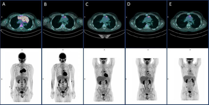
Misinterpretation of an inflammatory FDG uptake in a patient treated for Hodgkin lymphoma: a case report.
Hodgkin Lymphoma (HL) is a malignancy involving lymph nodes and lymphatic system. [18F]F-FDG PET/CT (FDG-PET) imaging is routinely used for staging, to assess early chemotherapy response (interim FDG-PET), at the end of treatment (EoT FDG-PET) and for the identification of disease recurrence. We present a case of a 39-year-old man treated for HL. FDG-PET scans performed after first line therapy (both Interim PET and at the end of therapy) demonstrated a persistent and significant mediastinal FDG uptake. The patient was treated with a second line therapy but the FDG-PET uptake did not change. After board discussion a new surgical, thoracoscopy-guided biopsy was performed. Histopathology demonstrated a dense fibrous tissue with occasional chronic inflammatory infiltrates. Persistent FDG-PET positivity may suggest refractory or relapsed disease. However, occasionally, non-malignant conditions are responsible for a persistent FDG uptake, not related to primary disease. An accurate evaluation of clinical history and previous imaging exams is mandatory for clinicians and others experts to avoid misinterpretations of FDG-PET results. Nevertheless, in some cases, only a more invasive procedure, such as a biopsy, may finally lead to a definitive diagnosis.

 求助内容:
求助内容: 应助结果提醒方式:


