The utility of a portable muscle ultrasound in the assessment of muscle alterations in children with acute lymphoblastic leukaemia

IF 8.9 1区 医学
Emma J. Verwaaijen, Annelienke M. van Hulst, Jeroen Molinger, Annelies Hartman, Rob Pieters, Martha A. Grootenhuis, Erica L.T. van den Akker, Marry M. van den Heuvel-Eibrink
{"title":"The utility of a portable muscle ultrasound in the assessment of muscle alterations in children with acute lymphoblastic leukaemia","authors":"Emma J. Verwaaijen,&nbsp;Annelienke M. van Hulst,&nbsp;Jeroen Molinger,&nbsp;Annelies Hartman,&nbsp;Rob Pieters,&nbsp;Martha A. Grootenhuis,&nbsp;Erica L.T. van den Akker,&nbsp;Marry M. van den Heuvel-Eibrink","doi":"10.1002/jcsm.13305","DOIUrl":null,"url":null,"abstract":"<div>\n \n \n <section>\n \n <h3> Background</h3>\n \n <p>During treatment for acute lymphoblastic leukaemia (ALL), children are prone to musculoskeletal deterioration. However, non-invasive tools to measure muscle mass and intramuscular alterations are limited. In this study we explored the feasibility of muscle ultrasound in children with ALL. Additionally, we analysed whether automated ultrasound outcomes of muscle size and intramuscular fat infiltration (IMAT) were associated with appendicular skeletal muscle mass (ASMM), muscle strength and physical performance.</p>\n </section>\n \n <section>\n \n <h3> Methods</h3>\n \n <p>Children with ALL, aged 3–18 years were included during maintenance therapy. Bilateral images of the rectus femoris muscle were captured using a portable linear array transducer connected to a tablet. Subsequently, an automated image annotation software (MuscleSound) was used to estimate cross-sectional area, muscle thickness and IMAT. Feasibility was assessed using acceptance (percentage of children approached who were enrolled), practicality (percentage of children that completed the ultrasound measurement after enrolment) and implementation (percentage of children that had sufficient imaging to be processed and analysed by the software). Assessments of ASMM by bioimpedance analysis, muscle strength using handheld dynamometry and timed physical performance tests were administered at the same visit. Multivariable linear models were estimated to study the associations between muscle ultrasound outcomes and ASMM, strength and physical performance, adjusted for sex, age, body mass index and ALL treatment week.</p>\n </section>\n \n <section>\n \n <h3> Results</h3>\n \n <p>Muscle ultrasound was performed in 60 out of 73 invited patients (76.9%), of which 37 were boys (61.7%), and median age was 6.1 years (range: 3–18.8 years). The acceptance was 98.7%, practicality 77.9% and implementation was 100%. Patients who refused the examination (<i>n</i> = 13) were younger (median: 3.6, range: 3–11.2 years) compared with the 60 examined children (<i>P</i> = 0.0009). In multivariable models, cross-sectional area was associated with ASMM (β = 0.49 <i>Z</i>-score, 95% confidence interval [CI]:0.3,2.4), knee-extension strength (β = 16.9 Newton [N], 95% CI: 4.8, 28.9), walking performance (β = −0.46 s, 95% CI: −0.75, −0.18) and rising from the floor (β = −1.07 s, 95% CI: −1.71, −0.42). Muscle thickness was associated with ASMM (β = 0.14 <i>Z</i>-score, 95% CI: 0.04, 0.24), knee-extension strength (β = 4.73 N, 95% CI: 0.99, 8.47), walking performance (β = −0.13 s, 95% CI: −0.22, −0.04) and rising from the floor (β = −0.28 s, 95% CI: −0.48, −0.08). IMAT was associated with knee-extension strength (β = −6.84 N, 95% CI: −12.26, −1.41), walking performance (β = 0.2 s, 95% CI: 0.08, 0.32) and rising from the floor (β = 0.54 s, 95% CI: 0.27, 0.8). None of the muscle ultrasound outcomes was associated with handgrip strength.</p>\n </section>\n \n <section>\n \n <h3> Conclusions</h3>\n \n <p>Portable muscle ultrasound appears a feasible and useful tool to measure muscle size and intramuscular alterations in children with ALL. Validation studies using magnetic resonance imaging (gold standard) are necessary to confirm accuracy in paediatric populations.</p>\n </section>\n </div>","PeriodicalId":186,"journal":{"name":"Journal of Cachexia, Sarcopenia and Muscle","volume":null,"pages":null},"PeriodicalIF":8.9000,"publicationDate":"2023-08-10","publicationTypes":"Journal Article","fieldsOfStudy":null,"isOpenAccess":false,"openAccessPdf":"https://onlinelibrary.wiley.com/doi/epdf/10.1002/jcsm.13305","citationCount":"0","resultStr":null,"platform":"Semanticscholar","paperid":null,"PeriodicalName":"Journal of Cachexia, Sarcopenia and Muscle","FirstCategoryId":"3","ListUrlMain":"https://onlinelibrary.wiley.com/doi/10.1002/jcsm.13305","RegionNum":1,"RegionCategory":"医学","ArticlePicture":[],"TitleCN":null,"AbstractTextCN":null,"PMCID":null,"EPubDate":"","PubModel":"","JCR":"","JCRName":"","Score":null,"Total":0}
引用次数: 0

Abstract

Background

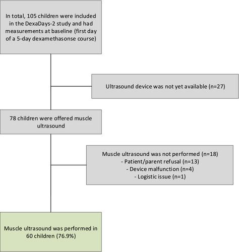
During treatment for acute lymphoblastic leukaemia (ALL), children are prone to musculoskeletal deterioration. However, non-invasive tools to measure muscle mass and intramuscular alterations are limited. In this study we explored the feasibility of muscle ultrasound in children with ALL. Additionally, we analysed whether automated ultrasound outcomes of muscle size and intramuscular fat infiltration (IMAT) were associated with appendicular skeletal muscle mass (ASMM), muscle strength and physical performance.

Methods

Children with ALL, aged 3–18 years were included during maintenance therapy. Bilateral images of the rectus femoris muscle were captured using a portable linear array transducer connected to a tablet. Subsequently, an automated image annotation software (MuscleSound) was used to estimate cross-sectional area, muscle thickness and IMAT. Feasibility was assessed using acceptance (percentage of children approached who were enrolled), practicality (percentage of children that completed the ultrasound measurement after enrolment) and implementation (percentage of children that had sufficient imaging to be processed and analysed by the software). Assessments of ASMM by bioimpedance analysis, muscle strength using handheld dynamometry and timed physical performance tests were administered at the same visit. Multivariable linear models were estimated to study the associations between muscle ultrasound outcomes and ASMM, strength and physical performance, adjusted for sex, age, body mass index and ALL treatment week.

Results

Muscle ultrasound was performed in 60 out of 73 invited patients (76.9%), of which 37 were boys (61.7%), and median age was 6.1 years (range: 3–18.8 years). The acceptance was 98.7%, practicality 77.9% and implementation was 100%. Patients who refused the examination (n = 13) were younger (median: 3.6, range: 3–11.2 years) compared with the 60 examined children (P = 0.0009). In multivariable models, cross-sectional area was associated with ASMM (β = 0.49 Z-score, 95% confidence interval [CI]:0.3,2.4), knee-extension strength (β = 16.9 Newton [N], 95% CI: 4.8, 28.9), walking performance (β = −0.46 s, 95% CI: −0.75, −0.18) and rising from the floor (β = −1.07 s, 95% CI: −1.71, −0.42). Muscle thickness was associated with ASMM (β = 0.14 Z-score, 95% CI: 0.04, 0.24), knee-extension strength (β = 4.73 N, 95% CI: 0.99, 8.47), walking performance (β = −0.13 s, 95% CI: −0.22, −0.04) and rising from the floor (β = −0.28 s, 95% CI: −0.48, −0.08). IMAT was associated with knee-extension strength (β = −6.84 N, 95% CI: −12.26, −1.41), walking performance (β = 0.2 s, 95% CI: 0.08, 0.32) and rising from the floor (β = 0.54 s, 95% CI: 0.27, 0.8). None of the muscle ultrasound outcomes was associated with handgrip strength.

Conclusions

Portable muscle ultrasound appears a feasible and useful tool to measure muscle size and intramuscular alterations in children with ALL. Validation studies using magnetic resonance imaging (gold standard) are necessary to confirm accuracy in paediatric populations.
Abstract Image

便携式肌肉超声在评估急性淋巴细胞白血病儿童肌肉改变中的作用。
背景:在急性淋巴细胞白血病(ALL)的治疗过程中,儿童的肌肉骨骼容易退化。然而,测量肌肉质量和肌内变化的非侵入性工具是有限的。在这项研究中,我们探讨了肌肉超声在ALL儿童中的可行性。此外,我们分析了肌肉大小和肌内脂肪浸润(IMAT)的自动超声结果是否与阑尾骨骼肌质量(ASMM)、肌肉力量和身体表现有关。方法:将3-18岁ALL患儿纳入维持治疗。使用连接到平板电脑的便携式线性阵列换能器拍摄股直肌的双侧图像。随后,使用自动图像注释软件(MuscleSound)来估计横截面积、肌肉厚度和IMAT。可行性评估采用接受度(被招募儿童的百分比)、实用性(在招募后完成超声波测量的儿童的比例)和实施度(有足够的图像可由软件处理和分析的儿童的比率)。在同一次访视中,通过生物阻抗分析评估ASMM,使用手持式测力仪评估肌肉力量,并进行定时身体性能测试。估计了多变量线性模型,以研究肌肉超声结果与ASMM、力量和身体表现之间的关系,并根据性别、年龄、体重指数和ALL治疗周进行了调整。结果:73名受邀患者中有60名(76.9%)进行了肌肉超声检查,其中37名为男孩(61.7%),中位年龄为6.1岁(范围:3-18.8岁)。接受率为98.7%,实用性为77.9%,实施率为100%。与60名接受检查的儿童(P=0.0009)相比,拒绝检查的患者(n=13)更年轻(中位数:3.6,范围:3-11.2岁)。在多变量模型中,截面积与ASMM(β=0.49 Z-评分,95%置信区间[CI]:0.3,2.4)、膝关节伸展强度(β=116.9 Newton[n],95%CI:4.8,28.9)、,肌肉厚度与ASMM(β=0.14Z-score,95%CI:0.04,0.24)、伸膝强度(β=4.73N,95%CI:0.99,8.47)、,行走性能(β=0.13s,95%可信区间:-0.22,-0.04)和从地板上爬起(β=0.28s,95%置信区间:-0.48,-0.08),行走性能(β=0.2s,95%CI:0.08,0.32)和从地板上站起来(β=0.54s,95%CI:0.27,0.8)。肌肉超声结果均与握力无关。结论:便携式肌肉超声是测量ALL儿童肌肉大小和肌肉改变的一种可行和有用的工具。使用磁共振成像(金标准)的验证研究对于确认儿科人群的准确性是必要的。
本文章由计算机程序翻译,如有差异,请以英文原文为准。
求助全文
约1分钟内获得全文 求助全文
来源期刊
Journal of Cachexia, Sarcopenia and Muscle
Journal of Cachexia, Sarcopenia and Muscle Medicine-Orthopedics and Sports Medicine
自引率
12.40%
发文量
0
期刊介绍: The Journal of Cachexia, Sarcopenia, and Muscle is a prestigious, peer-reviewed international publication committed to disseminating research and clinical insights pertaining to cachexia, sarcopenia, body composition, and the physiological and pathophysiological alterations occurring throughout the lifespan and in various illnesses across the spectrum of life sciences. This journal serves as a valuable resource for physicians, biochemists, biologists, dieticians, pharmacologists, and students alike.
×
引用
GB/T 7714-2015
复制
MLA
复制
APA
复制
导出至
BibTeX EndNote RefMan NoteFirst NoteExpress
×
提示
您的信息不完整,为了账户安全,请先补充。
现在去补充
×
提示
您因"违规操作"
具体请查看互助需知
我知道了
×
提示
确定
请完成安全验证×
copy
已复制链接
快去分享给好友吧!
我知道了
右上角分享
点击右上角分享
0
联系我们:info@booksci.cn Book学术提供免费学术资源搜索服务,方便国内外学者检索中英文文献。致力于提供最便捷和优质的服务体验。 Copyright © 2023 布克学术 All rights reserved.
京ICP备2023020795号-1
ghs 京公网安备 11010802042870号
Book学术文献互助
Book学术文献互助群
群 号:481959085
Book学术官方微信