Yi Dong, Wen-Ping Wang, Andre Ignee, Dan Zuo, Yi-Jie Qiu, Qi Zhang, Xiu-Yun Lu, Sheng Chen, Christoph Frank Dietrich
{"title":"The diagnostic value of Doppler Resistive Index in the differential diagnosis of focal liver lesions.","authors":"Yi Dong, Wen-Ping Wang, Andre Ignee, Dan Zuo, Yi-Jie Qiu, Qi Zhang, Xiu-Yun Lu, Sheng Chen, Christoph Frank Dietrich","doi":"10.15557/jou.2023.0010","DOIUrl":null,"url":null,"abstract":"<p><strong>Aim: </strong>To investigate the diagnostic value of resistance index (RI) in differentiating focal liver lesions.</p><p><strong>Patients and methods: </strong>In this retrospective study, a total of 576 patients with histologically confirmed focal liver lesions were included. Each patient underwent B-mode ultrasound examination and color Doppler ultrasound examination. The RI values of different focal liver lesions were recorded and compared.</p><p><strong>Results: </strong>The mean RI value of benign lesions was significantly lower than that of malignant lesions (0.54 ± 0.10 <i>vs</i>. 0.71 ± 0.12) (<i>p</i> <0.05). In malignant lesions, the RI value of intrahepatic cholangiocarcinoma was significantly lower than that of hepatocellular carcinoma lesions. Furthermore, in hepatocellular carcinoma lesions, the RI of large lesions (group 4: >10 cm) was significantly lower than that of small lesions (group 1: ≤2 cm, group 2: 2-5 cm) (<i>p</i> <0.05). Taken RI of 0.615 as a cutoff value to differentiate malignant and benign lesions, the sensitivity, specificity, positive predictive value and negative predictive value were 82.80%, 81.00%, 81.34% and 82.48%, respectively.</p><p><strong>Conclusion: </strong>Color Doppler ultrasound examination is a valuable imaging method in detecting blood flow signal within liver lesions. The RI parameter should be helpful in differentiating malignant and benign liver tumors.</p>","PeriodicalId":45612,"journal":{"name":"Journal of Ultrasonography","volume":"23 93","pages":"e45-e52"},"PeriodicalIF":1.5000,"publicationDate":"2023-06-01","publicationTypes":"Journal Article","fieldsOfStudy":null,"isOpenAccess":false,"openAccessPdf":"https://ftp.ncbi.nlm.nih.gov/pub/pmc/oa_pdf/11/a6/jou-23-93-jou.2023.0010.PMC10379844.pdf","citationCount":"2","resultStr":null,"platform":"Semanticscholar","paperid":null,"PeriodicalName":"Journal of Ultrasonography","FirstCategoryId":"1085","ListUrlMain":"https://doi.org/10.15557/jou.2023.0010","RegionNum":0,"RegionCategory":null,"ArticlePicture":[],"TitleCN":null,"AbstractTextCN":null,"PMCID":null,"EPubDate":"","PubModel":"","JCR":"Q3","JCRName":"RADIOLOGY, NUCLEAR MEDICINE & MEDICAL IMAGING","Score":null,"Total":0}
引用次数: 2
Abstract
Aim: To investigate the diagnostic value of resistance index (RI) in differentiating focal liver lesions.
Patients and methods: In this retrospective study, a total of 576 patients with histologically confirmed focal liver lesions were included. Each patient underwent B-mode ultrasound examination and color Doppler ultrasound examination. The RI values of different focal liver lesions were recorded and compared.
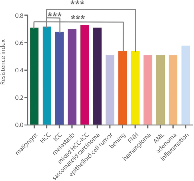
Results: The mean RI value of benign lesions was significantly lower than that of malignant lesions (0.54 ± 0.10 vs. 0.71 ± 0.12) (p <0.05). In malignant lesions, the RI value of intrahepatic cholangiocarcinoma was significantly lower than that of hepatocellular carcinoma lesions. Furthermore, in hepatocellular carcinoma lesions, the RI of large lesions (group 4: >10 cm) was significantly lower than that of small lesions (group 1: ≤2 cm, group 2: 2-5 cm) (p <0.05). Taken RI of 0.615 as a cutoff value to differentiate malignant and benign lesions, the sensitivity, specificity, positive predictive value and negative predictive value were 82.80%, 81.00%, 81.34% and 82.48%, respectively.
Conclusion: Color Doppler ultrasound examination is a valuable imaging method in detecting blood flow signal within liver lesions. The RI parameter should be helpful in differentiating malignant and benign liver tumors.

 求助内容:
求助内容: 应助结果提醒方式:


