Histopathological findings affect quantitative values of single photon emission computed tomography in patients with antiresorptive agent-related osteonecrosis of the jaws.
{"title":"Histopathological findings affect quantitative values of single photon emission computed tomography in patients with antiresorptive agent-related osteonecrosis of the jaws.","authors":"Taro Okui, Yoshikazu Kobayashi, Madoka Isomura, Masakazu Tsujimoto, Koji Satoh, Hiroshi Toyama","doi":"10.20407/fmj.2022-025","DOIUrl":null,"url":null,"abstract":"<p><strong>Objectives: </strong>This study investigated the relationships between quantitative values calculated from bone single photon emission computed tomography/computed tomography (SPECT/CT) images and histopathological findings observed in surgical specimens from patients with antiresorptive agent-related osteonecrosis of the jaw (ARONJ); it sought to clarify histopathological factors that cause accumulation in bone SPECT/CT images of patients with ARONJ.</p><p><strong>Methods: </strong>This study included 81 pathological specimens of 21 lesions obtained from 18 patients with ARONJ who underwent SPECT/CT and jaw resection. The maximum standardized uptake value (SUV<sub>max</sub>) of each volume of interest of the specimens was calculated using RAVAT<sup>®</sup> software. The ratio of the SUV<sub>max</sub> to the mean value of SUV<sub>max</sub> in temporal bone was termed rSUV<sub>max</sub>. The rSUV<sub>max</sub> and pathological findings (sequestration, degree of fibrosis, degree of trabecular bone destruction, degree of inflammatory cell infiltration, and vascularity) were compared using the Mann-Whitney U test and the Kruskal-Wallis test.</p><p><strong>Results: </strong>In univariate analysis with rSUV<sub>max</sub> as the dependent variable, the pathological findings of sequestration (P=0.058), degree of fibrosis (P=0.810), degree of trabecular bone destruction (P=0.237), degree of inflammatory cell infiltration (P=0.120), and vascularity (P=0.111) showed no significant difference among the groups for each variable.</p><p><strong>Conclusions: </strong>We found no association between quantitative values in bone SPECT/CT and histological changes in ARONJ, probably because bone SPECT/CT has limited spatial resolution. Limitations of this study may include the imaging findings of a decrease in tracer accumulation because of an involucrum of necrosed bone, various histopathological findings in ARONJ, and failure to consider the effect of preoperative anti-inflammatory treatment.</p>","PeriodicalId":33657,"journal":{"name":"Fujita Medical Journal","volume":"9 3","pages":"186-193"},"PeriodicalIF":0.0000,"publicationDate":"2023-08-01","publicationTypes":"Journal Article","fieldsOfStudy":null,"isOpenAccess":false,"openAccessPdf":"https://www.ncbi.nlm.nih.gov/pmc/articles/PMC10405893/pdf/","citationCount":"0","resultStr":null,"platform":"Semanticscholar","paperid":null,"PeriodicalName":"Fujita Medical Journal","FirstCategoryId":"1085","ListUrlMain":"https://doi.org/10.20407/fmj.2022-025","RegionNum":0,"RegionCategory":null,"ArticlePicture":[],"TitleCN":null,"AbstractTextCN":null,"PMCID":null,"EPubDate":"","PubModel":"","JCR":"","JCRName":"","Score":null,"Total":0}
引用次数: 0
Abstract
Objectives: This study investigated the relationships between quantitative values calculated from bone single photon emission computed tomography/computed tomography (SPECT/CT) images and histopathological findings observed in surgical specimens from patients with antiresorptive agent-related osteonecrosis of the jaw (ARONJ); it sought to clarify histopathological factors that cause accumulation in bone SPECT/CT images of patients with ARONJ.
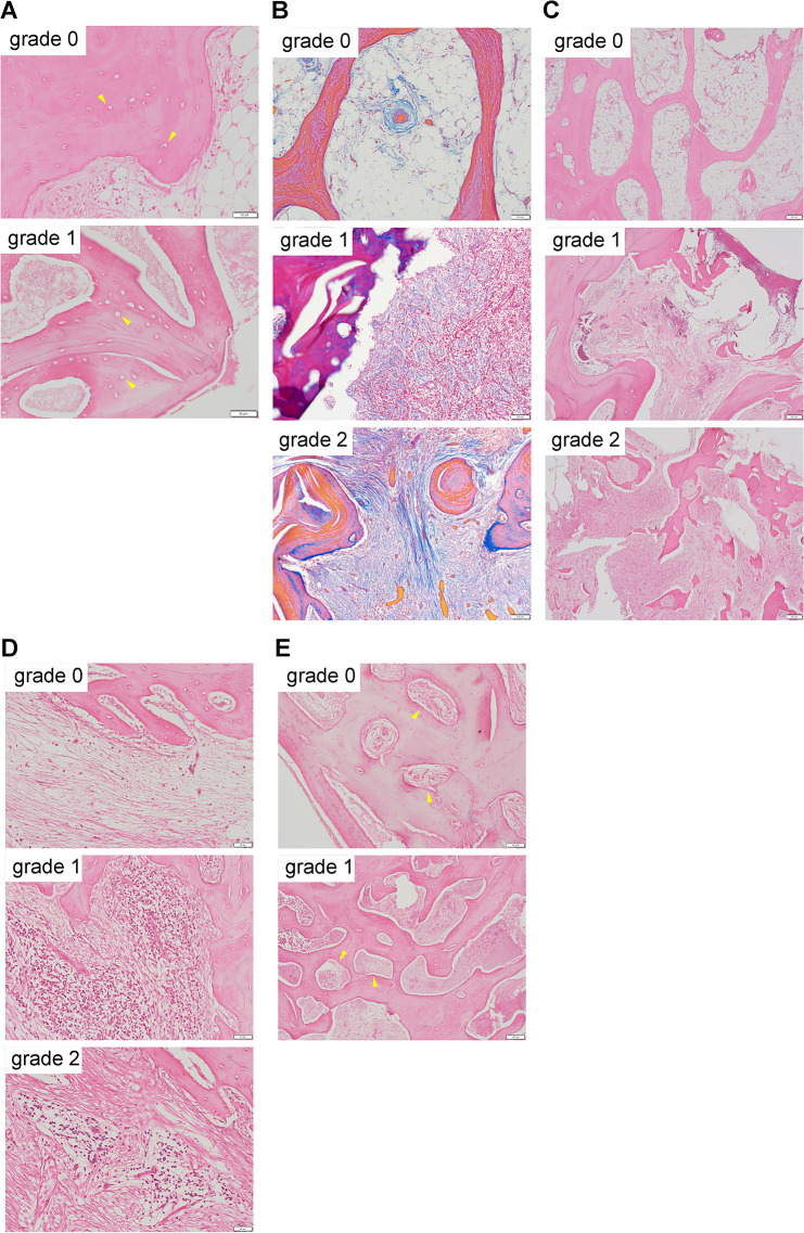
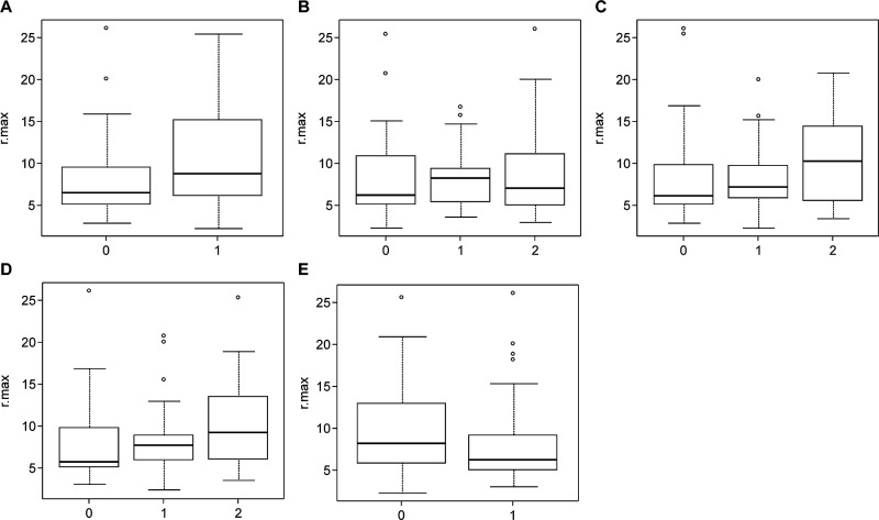
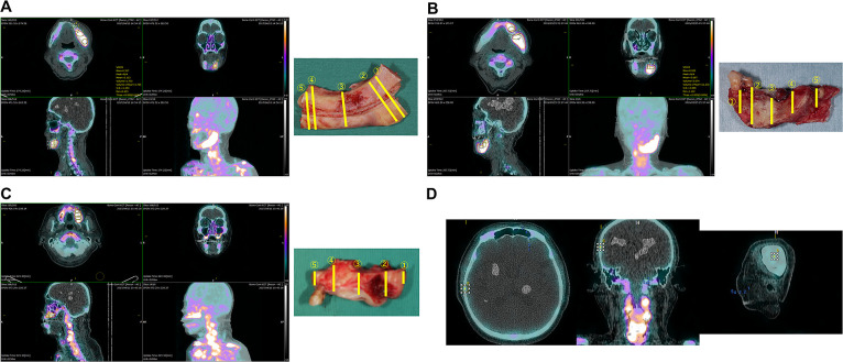
Methods: This study included 81 pathological specimens of 21 lesions obtained from 18 patients with ARONJ who underwent SPECT/CT and jaw resection. The maximum standardized uptake value (SUVmax) of each volume of interest of the specimens was calculated using RAVAT® software. The ratio of the SUVmax to the mean value of SUVmax in temporal bone was termed rSUVmax. The rSUVmax and pathological findings (sequestration, degree of fibrosis, degree of trabecular bone destruction, degree of inflammatory cell infiltration, and vascularity) were compared using the Mann-Whitney U test and the Kruskal-Wallis test.
Results: In univariate analysis with rSUVmax as the dependent variable, the pathological findings of sequestration (P=0.058), degree of fibrosis (P=0.810), degree of trabecular bone destruction (P=0.237), degree of inflammatory cell infiltration (P=0.120), and vascularity (P=0.111) showed no significant difference among the groups for each variable.
Conclusions: We found no association between quantitative values in bone SPECT/CT and histological changes in ARONJ, probably because bone SPECT/CT has limited spatial resolution. Limitations of this study may include the imaging findings of a decrease in tracer accumulation because of an involucrum of necrosed bone, various histopathological findings in ARONJ, and failure to consider the effect of preoperative anti-inflammatory treatment.

 求助内容:
求助内容: 应助结果提醒方式:


