Si-Ping Luo, Han-Wen Zhang, Yi Lei, Yu-Ning Feng, Juan Yu, Fan Lin
{"title":"Transient partial regression of intracranial germ cell tumor in adult thalamus: A case report.","authors":"Si-Ping Luo, Han-Wen Zhang, Yi Lei, Yu-Ning Feng, Juan Yu, Fan Lin","doi":"10.3389/fradi.2022.781475","DOIUrl":null,"url":null,"abstract":"<p><strong>Background: </strong>Intracranial germ cell tumors (GCTs) are a relatively rare malignancy in clinical practice. Natural regression of this tumor is also uncommon. We describe a rare case of an intracranial GCT in the thalamus of an adult that showed spontaneous regression and recurrence after steroid therapy.</p><p><strong>Case description: </strong>A 38-year-old male patient's MRI of the head suggested space-occupying masses in the left thalamus and midbrain. MRI examination revealed demyelination or granulomatous lesions. After high dose steroid treatment, the symptoms improved. The lesions were significantly reduced on repeat MRI, and oral steroid therapy was continued after discharge. The patient's symptoms deteriorated 1 month prior to a re-examination with head MRI, which revealed that the mass within the intracranial space was larger than on the previous image. He revisited the Department of Neurosurgery of our hospital and underwent left thalamic/pontine mass resection on October 16, 2019, and the pathological results showed that the tumor was a GCT.</p><p><strong>Conclusion: </strong>Intracranial GCTs are rare in the adult thalamus but should be considered in the differential diagnosis. The intracranial GCT regression seen in this case may be a short-lived phenomenon arising from complex immune responses caused by the intervention.</p>","PeriodicalId":73101,"journal":{"name":"Frontiers in radiology","volume":"2 ","pages":"781475"},"PeriodicalIF":0.0000,"publicationDate":"2022-01-01","publicationTypes":"Journal Article","fieldsOfStudy":null,"isOpenAccess":false,"openAccessPdf":"https://www.ncbi.nlm.nih.gov/pmc/articles/PMC10365112/pdf/","citationCount":"0","resultStr":null,"platform":"Semanticscholar","paperid":null,"PeriodicalName":"Frontiers in radiology","FirstCategoryId":"1085","ListUrlMain":"https://doi.org/10.3389/fradi.2022.781475","RegionNum":0,"RegionCategory":null,"ArticlePicture":[],"TitleCN":null,"AbstractTextCN":null,"PMCID":null,"EPubDate":"","PubModel":"","JCR":"","JCRName":"","Score":null,"Total":0}
引用次数: 0
Abstract
Background: Intracranial germ cell tumors (GCTs) are a relatively rare malignancy in clinical practice. Natural regression of this tumor is also uncommon. We describe a rare case of an intracranial GCT in the thalamus of an adult that showed spontaneous regression and recurrence after steroid therapy.
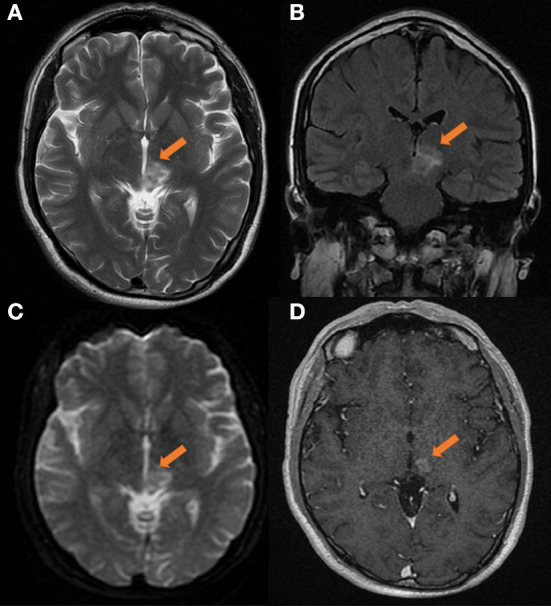
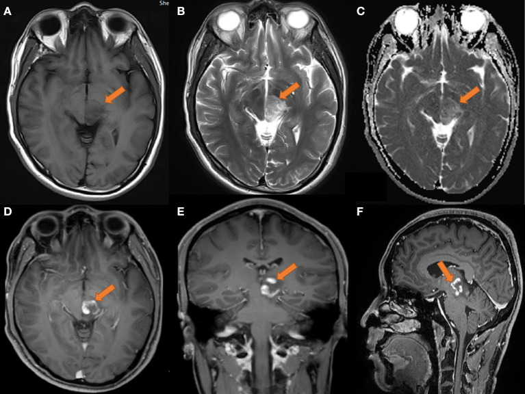
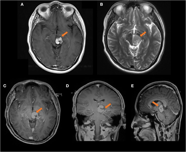
Case description: A 38-year-old male patient's MRI of the head suggested space-occupying masses in the left thalamus and midbrain. MRI examination revealed demyelination or granulomatous lesions. After high dose steroid treatment, the symptoms improved. The lesions were significantly reduced on repeat MRI, and oral steroid therapy was continued after discharge. The patient's symptoms deteriorated 1 month prior to a re-examination with head MRI, which revealed that the mass within the intracranial space was larger than on the previous image. He revisited the Department of Neurosurgery of our hospital and underwent left thalamic/pontine mass resection on October 16, 2019, and the pathological results showed that the tumor was a GCT.
Conclusion: Intracranial GCTs are rare in the adult thalamus but should be considered in the differential diagnosis. The intracranial GCT regression seen in this case may be a short-lived phenomenon arising from complex immune responses caused by the intervention.

 求助内容:
求助内容: 应助结果提醒方式:


