{"title":"Ex vivo cultivated retinal pigment epithelial cell transplantation for the treatment of rabbit corneal endothelial dysfunction.","authors":"Chunxiao Dong, Dulei Zou, Haoyun Duan, Xiangyue Hu, Qingjun Zhou, Weiyun Shi, Zongyi Li","doi":"10.1186/s40662-023-00351-4","DOIUrl":null,"url":null,"abstract":"<p><strong>Objective: </strong>Stem cell therapy is a promising strategy for the treatment of corneal endothelial dysfunction, and the need to find functional alternative seed cells of corneal endothelial cells (CECs) is urgent. Here, we determined the feasibility of using the retinal pigment epithelium (RPE) as an equivalent substitute for the treatment of corneal endothelial dysfunction.</p><p><strong>Methods: </strong>RPE cells and CECs in situ were obtained from healthy New Zealand male rabbits, and the similarities and differences between them were analyzed by electron microscopy, immunofluorescent staining, and quantitative real-time reverse transcription polymerase chain reaction (qRT-PCR). Rabbit primary RPE cells and CECs were isolated and cultivated ex vivo, and Na+/K+-ATPase activity and cellular permeability were detected at passage 2. The injection of cultivated rabbit primary RPE cells, CECs and human embryonic stem cell (hESC)-derived RPE cells was performed on rabbits with corneal endothelial dysfunction. Then, the therapeutic effects were evaluated by corneal transparency, central corneal thickness, enzyme linked immunosorbent assay (ELISA), qRT-PCR and immunofluorescent staining.</p><p><strong>Results: </strong>The rabbit RPE cells were similar in form to CECs in situ and ex vivo, showing a larger regular hexagonal shape and a lower cell density, with numerous tightly formed cell junctions and hemidesmosomes. Moreover, RPE cells presented a stronger barrier and ionic pumping capacity than CECs. When intracamerally injected into the rabbits, the transplanted primary RPE cells could dissolve corneal edema and decrease corneal thickness, with effects similar to those of CECs. In addition, the transplantation of hESC-derived RPE cells exhibited a similar therapeutic effect and restored corneal transparency and thickness within seven days. qRT-PCR results showed that the expressions of CEC markers, like CD200 and S100A4, increased, and the RPE markers OTX2, BEST1 and MITF significantly decreased in the transplanted RPE cells. Furthermore, we have demonstrated that rabbits transplanted with hESC-derived RPE cells maintained normal corneal thickness and exhibited slight pigmentation in the central cornea one month after surgery. Immunostaining results showed that the HuNu-positive transplanted cells survived and expressed ZO1, ATP1A1 and MITF.</p><p><strong>Conclusion: </strong>RPE cells and CECs showed high structural and functional similarities in barrier and pump characteristics. Intracameral injection of primary RPE cells and hESC-derived RPE cells can effectively restore rabbit corneal clarity and thickness and maintain normal corneal function. This study is the first to report the effectiveness of RPE cells for corneal endothelial dysfunction, suggesting the feasibility of hESC-derived RPE cells as an equivalent substitute for CECs.</p>","PeriodicalId":12194,"journal":{"name":"Eye and Vision","volume":null,"pages":null},"PeriodicalIF":4.1000,"publicationDate":"2023-08-02","publicationTypes":"Journal Article","fieldsOfStudy":null,"isOpenAccess":false,"openAccessPdf":"https://www.ncbi.nlm.nih.gov/pmc/articles/PMC10394777/pdf/","citationCount":"0","resultStr":null,"platform":"Semanticscholar","paperid":null,"PeriodicalName":"Eye and Vision","FirstCategoryId":"3","ListUrlMain":"https://doi.org/10.1186/s40662-023-00351-4","RegionNum":1,"RegionCategory":"医学","ArticlePicture":[],"TitleCN":null,"AbstractTextCN":null,"PMCID":null,"EPubDate":"","PubModel":"","JCR":"Q1","JCRName":"OPHTHALMOLOGY","Score":null,"Total":0}
引用次数: 0
Abstract
Objective: Stem cell therapy is a promising strategy for the treatment of corneal endothelial dysfunction, and the need to find functional alternative seed cells of corneal endothelial cells (CECs) is urgent. Here, we determined the feasibility of using the retinal pigment epithelium (RPE) as an equivalent substitute for the treatment of corneal endothelial dysfunction.
Methods: RPE cells and CECs in situ were obtained from healthy New Zealand male rabbits, and the similarities and differences between them were analyzed by electron microscopy, immunofluorescent staining, and quantitative real-time reverse transcription polymerase chain reaction (qRT-PCR). Rabbit primary RPE cells and CECs were isolated and cultivated ex vivo, and Na+/K+-ATPase activity and cellular permeability were detected at passage 2. The injection of cultivated rabbit primary RPE cells, CECs and human embryonic stem cell (hESC)-derived RPE cells was performed on rabbits with corneal endothelial dysfunction. Then, the therapeutic effects were evaluated by corneal transparency, central corneal thickness, enzyme linked immunosorbent assay (ELISA), qRT-PCR and immunofluorescent staining.
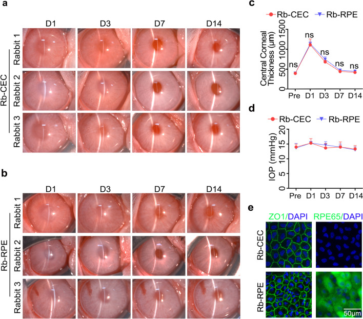
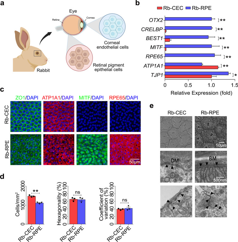
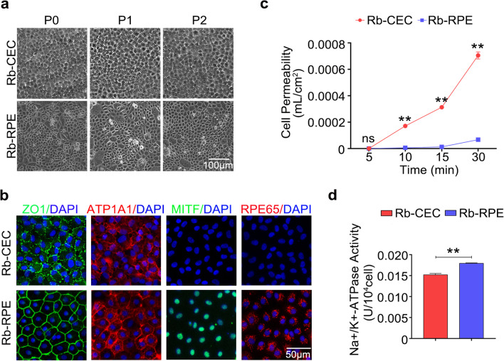
Results: The rabbit RPE cells were similar in form to CECs in situ and ex vivo, showing a larger regular hexagonal shape and a lower cell density, with numerous tightly formed cell junctions and hemidesmosomes. Moreover, RPE cells presented a stronger barrier and ionic pumping capacity than CECs. When intracamerally injected into the rabbits, the transplanted primary RPE cells could dissolve corneal edema and decrease corneal thickness, with effects similar to those of CECs. In addition, the transplantation of hESC-derived RPE cells exhibited a similar therapeutic effect and restored corneal transparency and thickness within seven days. qRT-PCR results showed that the expressions of CEC markers, like CD200 and S100A4, increased, and the RPE markers OTX2, BEST1 and MITF significantly decreased in the transplanted RPE cells. Furthermore, we have demonstrated that rabbits transplanted with hESC-derived RPE cells maintained normal corneal thickness and exhibited slight pigmentation in the central cornea one month after surgery. Immunostaining results showed that the HuNu-positive transplanted cells survived and expressed ZO1, ATP1A1 and MITF.
Conclusion: RPE cells and CECs showed high structural and functional similarities in barrier and pump characteristics. Intracameral injection of primary RPE cells and hESC-derived RPE cells can effectively restore rabbit corneal clarity and thickness and maintain normal corneal function. This study is the first to report the effectiveness of RPE cells for corneal endothelial dysfunction, suggesting the feasibility of hESC-derived RPE cells as an equivalent substitute for CECs.
期刊介绍:
Eye and Vision is an open access, peer-reviewed journal for ophthalmologists and visual science specialists. It welcomes research articles, reviews, methodologies, commentaries, case reports, perspectives and short reports encompassing all aspects of eye and vision. Topics of interest include but are not limited to: current developments of theoretical, experimental and clinical investigations in ophthalmology, optometry and vision science which focus on novel and high-impact findings on central issues pertaining to biology, pathophysiology and etiology of eye diseases as well as advances in diagnostic techniques, surgical treatment, instrument updates, the latest drug findings, results of clinical trials and research findings. It aims to provide ophthalmologists and visual science specialists with the latest developments in theoretical, experimental and clinical investigations in eye and vision.
 求助内容:
求助内容: 应助结果提醒方式:


