{"title":"A case of chronic gastric anisakiasis coexisting with early gastric cancer.","authors":"Eiko Sakurai, Masaaki Okubo, Yutaka Tsutsumi, Tomoyuki Shibata, Tomomitsu Tahara, Yuka Kiriyama, Ayano Michiba, Naoki Ohmiya, Tetsuya Tsukamoto","doi":"10.20407/fmj.2022-010","DOIUrl":null,"url":null,"abstract":"<p><strong>Background: </strong>Anisakiasis is a parasitic disease caused by the consumption of raw or undercooked fish that is infected with <i>Anisakis</i> third-stage larvae. In countries, such as Japan, Italy, and Spain, where people have a custom of eating raw or marinated fish, anisakiasis is a common infection. Although anisakiasis has been reported in the gastrointestinal tract in several countries, reports of anisakiasis accompanied by cancer are rare.</p><p><strong>Case presentation: </strong>We present the rare case of a 40-year-old male patient with anisakiasis coexisting with mucosal gastric cancer. Submucosal gastric cancer was suspected on gastric endoscopy and endoscopic ultrasonography. After laparoscopic distal gastrectomy, granulomatous inflammation with <i>Anisakis</i> larvae in the submucosa was pathologically revealed beneath mucosal tubular adenocarcinoma. Histological and immunohistochemical investigation showed cancer cells as intestinal absorptive-type cells that did not produce mucin.</p><p><strong>Conclusion: </strong><i>Anisakis</i> larvae could have invaded the cancer cells selectively because of the lack of mucin in the cancerous epithelium. Anisakiasis coexisting with cancer is considered reasonable rather than coincidental. In cancer with anisakiasis, preoperative diagnosis may be difficult because anisakiasis leads to morphological changes in the cancer.</p>","PeriodicalId":33657,"journal":{"name":"Fujita Medical Journal","volume":"9 2","pages":"163-169"},"PeriodicalIF":0.0000,"publicationDate":"2023-05-01","publicationTypes":"Journal Article","fieldsOfStudy":null,"isOpenAccess":false,"openAccessPdf":"https://www.ncbi.nlm.nih.gov/pmc/articles/PMC10206891/pdf/","citationCount":"1","resultStr":null,"platform":"Semanticscholar","paperid":null,"PeriodicalName":"Fujita Medical Journal","FirstCategoryId":"1085","ListUrlMain":"https://doi.org/10.20407/fmj.2022-010","RegionNum":0,"RegionCategory":null,"ArticlePicture":[],"TitleCN":null,"AbstractTextCN":null,"PMCID":null,"EPubDate":"","PubModel":"","JCR":"","JCRName":"","Score":null,"Total":0}
引用次数: 1
Abstract
Background: Anisakiasis is a parasitic disease caused by the consumption of raw or undercooked fish that is infected with Anisakis third-stage larvae. In countries, such as Japan, Italy, and Spain, where people have a custom of eating raw or marinated fish, anisakiasis is a common infection. Although anisakiasis has been reported in the gastrointestinal tract in several countries, reports of anisakiasis accompanied by cancer are rare.
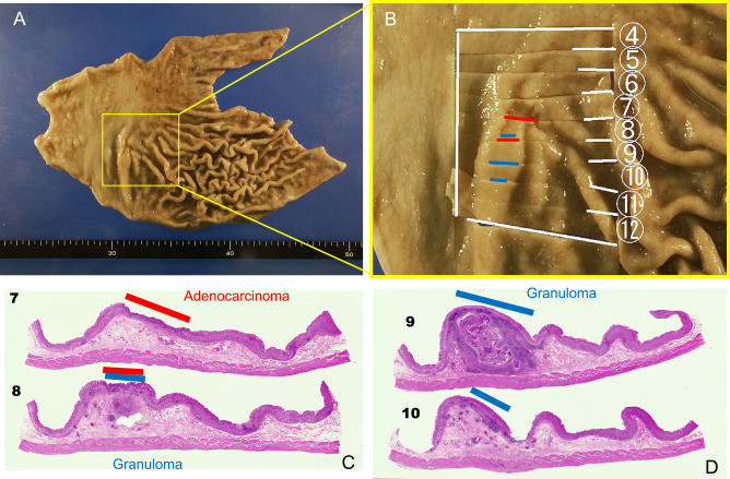
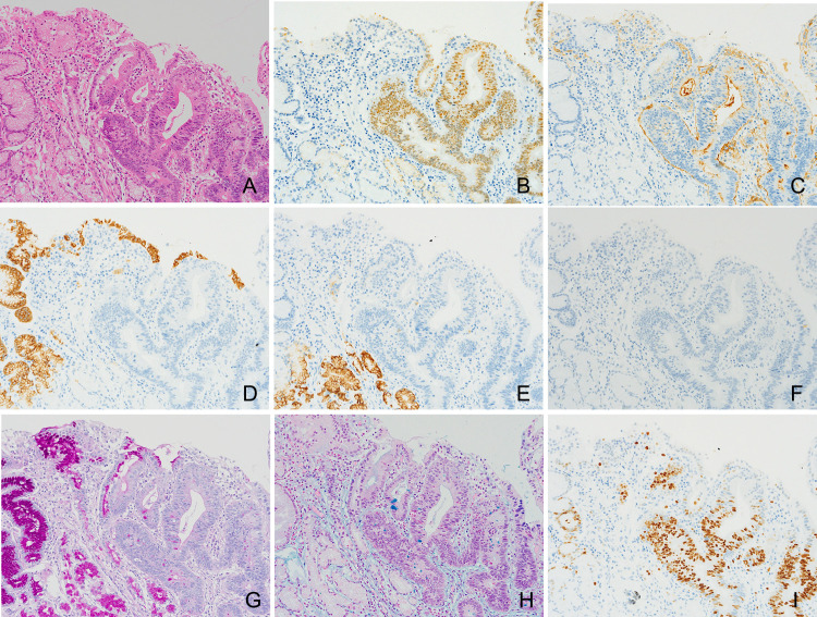
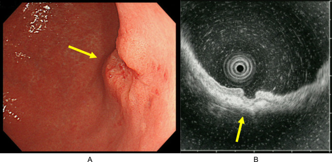
Case presentation: We present the rare case of a 40-year-old male patient with anisakiasis coexisting with mucosal gastric cancer. Submucosal gastric cancer was suspected on gastric endoscopy and endoscopic ultrasonography. After laparoscopic distal gastrectomy, granulomatous inflammation with Anisakis larvae in the submucosa was pathologically revealed beneath mucosal tubular adenocarcinoma. Histological and immunohistochemical investigation showed cancer cells as intestinal absorptive-type cells that did not produce mucin.
Conclusion: Anisakis larvae could have invaded the cancer cells selectively because of the lack of mucin in the cancerous epithelium. Anisakiasis coexisting with cancer is considered reasonable rather than coincidental. In cancer with anisakiasis, preoperative diagnosis may be difficult because anisakiasis leads to morphological changes in the cancer.

 求助内容:
求助内容: 应助结果提醒方式:


