{"title":"Importance of correcting alar base ptosis during primary cleft lip repair.","authors":"Maki Inukai, Yoshikazu Inoue, Yoshimi Sano, Satoko Onishi, Takayuki Okumoto, Ichiro Uyama","doi":"10.20407/fmj.2022-014","DOIUrl":null,"url":null,"abstract":"<p><strong>Objectives: </strong>Until 1999 at our hospital, primary cleft lip repair was performed by the straight-line method and external rhinoplasty was performed by the inverted trapezoidal suture method with bilateral reverse-U incisions for children with cleft lip and palate. Subsequently, repeated surgical corrections of the external nasal morphology became necessary during the growth period, often with unsatisfactory results because repeated external rhinoplasty results in a stronger scar contracture. From 2000 to 2004, we performed external rhinoplasty after patients had stopped growing; however, delaying surgery created a psychological burden for patients. Therefore, since 2005, we have focused on improving alar base ptosis and forming the nostril sill during the primary surgery. This study was performed to subjectively and objectively evaluate whether the current surgical method or the earlier technique produces a better treatment outcome.</p><p><strong>Methods: </strong>We subjectively and objectively evaluated alar base asymmetry after primary cleft lip repair but before bone grafting for alveolar cleft repair. For the objective evaluation, we measured the angle of alar base ptosis in frontal view photographs taken at the age of 6 or 7 years in patients who underwent repair before 1999 (Group A) and after 2005 (Group B).</p><p><strong>Results: </strong>The median angle was 2.75° in Group A and 1.50° in Group B, demonstrating a significant difference (P=0.04).</p><p><strong>Conclusions: </strong>The current surgical method, which reflects our focus on improving alar base ptosis and forming the nostril sill, subjectively and objectively improved the external nasal morphology.</p>","PeriodicalId":33657,"journal":{"name":"Fujita Medical Journal","volume":"9 2","pages":"121-125"},"PeriodicalIF":0.0000,"publicationDate":"2023-05-01","publicationTypes":"Journal Article","fieldsOfStudy":null,"isOpenAccess":false,"openAccessPdf":"https://www.ncbi.nlm.nih.gov/pmc/articles/PMC10206893/pdf/","citationCount":"0","resultStr":null,"platform":"Semanticscholar","paperid":null,"PeriodicalName":"Fujita Medical Journal","FirstCategoryId":"1085","ListUrlMain":"https://doi.org/10.20407/fmj.2022-014","RegionNum":0,"RegionCategory":null,"ArticlePicture":[],"TitleCN":null,"AbstractTextCN":null,"PMCID":null,"EPubDate":"","PubModel":"","JCR":"","JCRName":"","Score":null,"Total":0}
引用次数: 0
Abstract
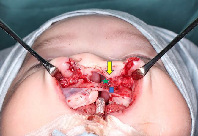
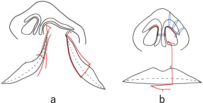
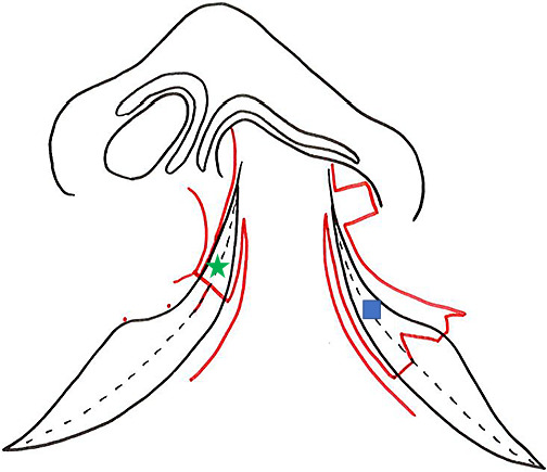
Objectives: Until 1999 at our hospital, primary cleft lip repair was performed by the straight-line method and external rhinoplasty was performed by the inverted trapezoidal suture method with bilateral reverse-U incisions for children with cleft lip and palate. Subsequently, repeated surgical corrections of the external nasal morphology became necessary during the growth period, often with unsatisfactory results because repeated external rhinoplasty results in a stronger scar contracture. From 2000 to 2004, we performed external rhinoplasty after patients had stopped growing; however, delaying surgery created a psychological burden for patients. Therefore, since 2005, we have focused on improving alar base ptosis and forming the nostril sill during the primary surgery. This study was performed to subjectively and objectively evaluate whether the current surgical method or the earlier technique produces a better treatment outcome.
Methods: We subjectively and objectively evaluated alar base asymmetry after primary cleft lip repair but before bone grafting for alveolar cleft repair. For the objective evaluation, we measured the angle of alar base ptosis in frontal view photographs taken at the age of 6 or 7 years in patients who underwent repair before 1999 (Group A) and after 2005 (Group B).
Results: The median angle was 2.75° in Group A and 1.50° in Group B, demonstrating a significant difference (P=0.04).
Conclusions: The current surgical method, which reflects our focus on improving alar base ptosis and forming the nostril sill, subjectively and objectively improved the external nasal morphology.

 求助内容:
求助内容: 应助结果提醒方式:


