Comparison of Image Quality and Radiation Dose Between Single-Energy and Dual-Energy Images for the Brain With Stereotactic Frames on Dual-Energy Cerebral CT.
Xiaojing Zhao, Wang Chao, Yi Shan, Jingkai Li, Cheng Zhao, Miao Zhang, Jie Lu
{"title":"Comparison of Image Quality and Radiation Dose Between Single-Energy and Dual-Energy Images for the Brain With Stereotactic Frames on Dual-Energy Cerebral CT.","authors":"Xiaojing Zhao, Wang Chao, Yi Shan, Jingkai Li, Cheng Zhao, Miao Zhang, Jie Lu","doi":"10.3389/fradi.2022.899100","DOIUrl":null,"url":null,"abstract":"<p><strong>Background: </strong>Preoperative stereotactic planning of deep brain stimulation (DBS) using computed tomography (CT) imaging in patients with Parkinson's disease (PD) is of clinical interest. However, frame-induced metal artifacts are common in clinical practice, which can be challenging for neurosurgeons to visualize brain structures.</p><p><strong>Objectives: </strong>To evaluate the image quality and radiation exposure of patients with stereotactic frame brain CT acquired using a dual-source CT (DSCT) system in single- and dual-energy modes.</p><p><strong>Materials and methods: </strong>We included 60 consecutive patients with Parkinson's disease (PD) and randomized them into two groups. CT images of the brain were performed using DSCT (Group A, an 80/Sn150 kVp dual-energy mode; Group B, a 120 kVp single-energy mode). One set of single-energy images (120 kVp) and 10 sets of virtual monochromatic images (50-140 keV) were obtained. Subjective image analysis of overall image quality was performed using a five-point Likert scale. For objective image quality evaluation, CT values, image noise, signal-to-noise ratio (SNR), and contrast-to-noise (CNR) were calculated. The radiation dose was recorded for each patient.</p><p><strong>Results: </strong>The mean effective radiation dose was reduced in the dual-energy mode (1.73 mSv ± 0.45 mSv) compared to the single-energy mode (3.16 mSv ± 0.64 mSv) (<i>p</i> < 0.001). Image noise was reduced by 46-52% for 120-140 keV VMI compared to 120 kVp images (both <i>p</i> < 0.01). CT values were higher at 100-140 keV than at 120 kVp images. At 120-140 keV, CT values of brain tissue showed significant differences at the level of the most severe metal artifacts (all <i>p</i> < 0.05). SNR was also higher in the dual-energy mode 90-140 keV compared to 120 kVp images, showing a significant difference between the two groups at 120-140 keV (all <i>p</i> < 0.01). The CNR was significantly better in Group A for 60-140 keV VMI compared to Group B (both <i>p</i> < 0.001). The highest subjective image scores were found in the 120 keV images, while 110-140 keV images had significantly higher scores than 120 kVp images (all <i>p</i> < 0.05).</p><p><strong>Conclusion: </strong>DSCT images using dual-energy modes provide better objective and subjective image quality for patients with PD at lower radiation doses compared to single-energy modes and facilitate brain tissue visualization with stereotactic frame DBS procedures.</p>","PeriodicalId":73101,"journal":{"name":"Frontiers in radiology","volume":"2 ","pages":"899100"},"PeriodicalIF":0.0000,"publicationDate":"2022-06-10","publicationTypes":"Journal Article","fieldsOfStudy":null,"isOpenAccess":false,"openAccessPdf":"https://www.ncbi.nlm.nih.gov/pmc/articles/PMC10364999/pdf/","citationCount":"0","resultStr":null,"platform":"Semanticscholar","paperid":null,"PeriodicalName":"Frontiers in radiology","FirstCategoryId":"1085","ListUrlMain":"https://doi.org/10.3389/fradi.2022.899100","RegionNum":0,"RegionCategory":null,"ArticlePicture":[],"TitleCN":null,"AbstractTextCN":null,"PMCID":null,"EPubDate":"2022/1/1 0:00:00","PubModel":"eCollection","JCR":"","JCRName":"","Score":null,"Total":0}
引用次数: 0
Abstract
Background: Preoperative stereotactic planning of deep brain stimulation (DBS) using computed tomography (CT) imaging in patients with Parkinson's disease (PD) is of clinical interest. However, frame-induced metal artifacts are common in clinical practice, which can be challenging for neurosurgeons to visualize brain structures.
Objectives: To evaluate the image quality and radiation exposure of patients with stereotactic frame brain CT acquired using a dual-source CT (DSCT) system in single- and dual-energy modes.
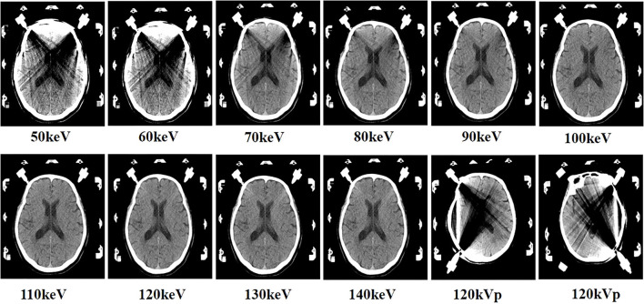
Materials and methods: We included 60 consecutive patients with Parkinson's disease (PD) and randomized them into two groups. CT images of the brain were performed using DSCT (Group A, an 80/Sn150 kVp dual-energy mode; Group B, a 120 kVp single-energy mode). One set of single-energy images (120 kVp) and 10 sets of virtual monochromatic images (50-140 keV) were obtained. Subjective image analysis of overall image quality was performed using a five-point Likert scale. For objective image quality evaluation, CT values, image noise, signal-to-noise ratio (SNR), and contrast-to-noise (CNR) were calculated. The radiation dose was recorded for each patient.
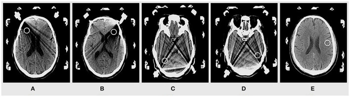
Results: The mean effective radiation dose was reduced in the dual-energy mode (1.73 mSv ± 0.45 mSv) compared to the single-energy mode (3.16 mSv ± 0.64 mSv) (p < 0.001). Image noise was reduced by 46-52% for 120-140 keV VMI compared to 120 kVp images (both p < 0.01). CT values were higher at 100-140 keV than at 120 kVp images. At 120-140 keV, CT values of brain tissue showed significant differences at the level of the most severe metal artifacts (all p < 0.05). SNR was also higher in the dual-energy mode 90-140 keV compared to 120 kVp images, showing a significant difference between the two groups at 120-140 keV (all p < 0.01). The CNR was significantly better in Group A for 60-140 keV VMI compared to Group B (both p < 0.001). The highest subjective image scores were found in the 120 keV images, while 110-140 keV images had significantly higher scores than 120 kVp images (all p < 0.05).
Conclusion: DSCT images using dual-energy modes provide better objective and subjective image quality for patients with PD at lower radiation doses compared to single-energy modes and facilitate brain tissue visualization with stereotactic frame DBS procedures.

 求助内容:
求助内容: 应助结果提醒方式:


