{"title":"Relevance maps: A weakly supervised segmentation method for 3D brain tumours in MRIs.","authors":"Sajith Rajapaksa, Farzad Khalvati","doi":"10.3389/fradi.2022.1061402","DOIUrl":null,"url":null,"abstract":"<p><p>With the increased reliance on medical imaging, Deep convolutional neural networks (CNNs) have become an essential tool in the medical imaging-based computer-aided diagnostic pipelines. However, training accurate and reliable classification models often require large fine-grained annotated datasets. To alleviate this, weakly-supervised methods can be used to obtain local information such as region of interest from global labels. This work proposes a weakly-supervised pipeline to extract Relevance Maps of medical images from pre-trained 3D classification models using localized perturbations. The extracted Relevance Map describes a given region's importance to the classification model and produces the segmentation for the region. Furthermore, we propose a novel optimal perturbation generation method that exploits 3D superpixels to find the most relevant area for a given classification using U-net architecture. This model is trained with perturbation loss, which maximizes the difference between unperturbed and perturbed predictions. We validated the effectiveness of our methodology by applying it to the segmentation of Glioma brain tumours in MRI scans using only classification labels for glioma type. The proposed method outperforms existing methods in both Dice Similarity Coefficient for segmentation and resolution for visualizations.</p>","PeriodicalId":73101,"journal":{"name":"Frontiers in radiology","volume":null,"pages":null},"PeriodicalIF":0.0000,"publicationDate":"2022-01-01","publicationTypes":"Journal Article","fieldsOfStudy":null,"isOpenAccess":false,"openAccessPdf":"https://www.ncbi.nlm.nih.gov/pmc/articles/PMC10365288/pdf/","citationCount":"0","resultStr":null,"platform":"Semanticscholar","paperid":null,"PeriodicalName":"Frontiers in radiology","FirstCategoryId":"1085","ListUrlMain":"https://doi.org/10.3389/fradi.2022.1061402","RegionNum":0,"RegionCategory":null,"ArticlePicture":[],"TitleCN":null,"AbstractTextCN":null,"PMCID":null,"EPubDate":"","PubModel":"","JCR":"","JCRName":"","Score":null,"Total":0}
引用次数: 0
Abstract
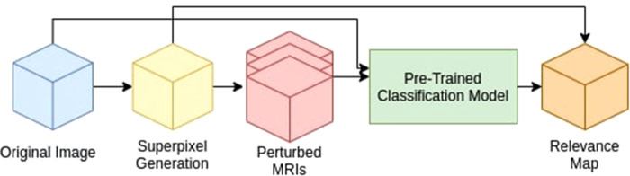
With the increased reliance on medical imaging, Deep convolutional neural networks (CNNs) have become an essential tool in the medical imaging-based computer-aided diagnostic pipelines. However, training accurate and reliable classification models often require large fine-grained annotated datasets. To alleviate this, weakly-supervised methods can be used to obtain local information such as region of interest from global labels. This work proposes a weakly-supervised pipeline to extract Relevance Maps of medical images from pre-trained 3D classification models using localized perturbations. The extracted Relevance Map describes a given region's importance to the classification model and produces the segmentation for the region. Furthermore, we propose a novel optimal perturbation generation method that exploits 3D superpixels to find the most relevant area for a given classification using U-net architecture. This model is trained with perturbation loss, which maximizes the difference between unperturbed and perturbed predictions. We validated the effectiveness of our methodology by applying it to the segmentation of Glioma brain tumours in MRI scans using only classification labels for glioma type. The proposed method outperforms existing methods in both Dice Similarity Coefficient for segmentation and resolution for visualizations.


 求助内容:
求助内容: 应助结果提醒方式:


