{"title":"Evolution of magnetic resonance imaging features in cerebral parenchyma from prolonged focal status epilepticus: a case study.","authors":"Sung Chul Lim, Jung Hee Cho, Young-Min Shon","doi":"10.47936/encephalitis.2021.00171","DOIUrl":null,"url":null,"abstract":"<p><p>It has rarely been documented that permanent alteration of cerebral structures occurs by focal status epilepticus (FSE). We report the case of a 16-year-old boy with FSE in whom serial T1-weighted magnetic resonance volumetry and conventional magnetic resonance imaging were useful for investigating an evolving pattern of morphological changes during and after the FSE, including cortical laminar necrosis (CLN), increased T2 signal intensities, and marked regional atrophy on the corresponding areas. Despite cessation of FSE after adequate medication (combination therapy including clobazam of 1 mg/kg/day), further significant cerebral atrophy was detected at the limited regions where discrete CLN had occurred during the FSE.</p>","PeriodicalId":72904,"journal":{"name":"Encephalitis (Seoul, Korea)","volume":"2 2","pages":"58-63"},"PeriodicalIF":0.0000,"publicationDate":"2022-04-01","publicationTypes":"Journal Article","fieldsOfStudy":null,"isOpenAccess":false,"openAccessPdf":"https://ftp.ncbi.nlm.nih.gov/pub/pmc/oa_pdf/35/78/encephalitis-2021-00171.PMC10295910.pdf","citationCount":"0","resultStr":null,"platform":"Semanticscholar","paperid":null,"PeriodicalName":"Encephalitis (Seoul, Korea)","FirstCategoryId":"1085","ListUrlMain":"https://doi.org/10.47936/encephalitis.2021.00171","RegionNum":0,"RegionCategory":null,"ArticlePicture":[],"TitleCN":null,"AbstractTextCN":null,"PMCID":null,"EPubDate":"","PubModel":"","JCR":"","JCRName":"","Score":null,"Total":0}
引用次数: 0
Abstract
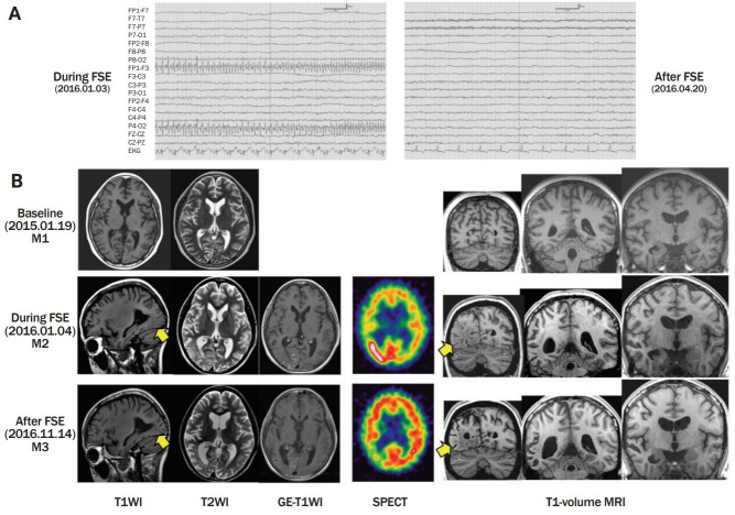
It has rarely been documented that permanent alteration of cerebral structures occurs by focal status epilepticus (FSE). We report the case of a 16-year-old boy with FSE in whom serial T1-weighted magnetic resonance volumetry and conventional magnetic resonance imaging were useful for investigating an evolving pattern of morphological changes during and after the FSE, including cortical laminar necrosis (CLN), increased T2 signal intensities, and marked regional atrophy on the corresponding areas. Despite cessation of FSE after adequate medication (combination therapy including clobazam of 1 mg/kg/day), further significant cerebral atrophy was detected at the limited regions where discrete CLN had occurred during the FSE.

 求助内容:
求助内容: 应助结果提醒方式:


