{"title":"Acute brainstem encephalitis associated with <i>Mycoplasma pneumoniae</i> in an adult: a case report.","authors":"Min-Hee Woo, Jung-Won Shin","doi":"10.47936/encephalitis.2021.00101","DOIUrl":null,"url":null,"abstract":"<p><p>Brainstem encephalitis (BE) associated with <i>Mycoplasma pneumoniae</i> in adults is rare, and the diagnosis is challenging. We describe an uncommon case of BE in an immunocompetent patient. A 43-year-old, otherwise healthy woman visited our emergency department with high fever and a sore throat, and 3 days later she returned with an altered drowsy mental status. Magnetic resonance imaging displayed diffuse swelling in bilateral cerebral regions involving the bilateral pons. The sera tested positive for the immunoglobulin (Ig) M antibody against <i>M. Pneumoniae</i> as detected by an enzyme immunoassay (EIA), and on hospital day 10, the level of IgM index against <i>M. pneumoniae</i> further increased from 1.5 to 2.1. We changed the antibiotic regimen from vancomycin and ceftriaxone to clarithromycin based on detection of <i>M. pneumoniae</i>, and we added intravenous immunoglobulin. After one month, the patient fully recovered from the neurological deficits. A follow-up brain magnetic resonance imaging was performed, which showed completely resolved lesions. Particle agglutination assay (PA) and EIA are both largely used to diagnose <i>M. pneumoniae</i>. Compared to the PA test, the EIA test could be a reliable tool because it separately measures IgM and IgG antibodies. We diagnosed BE associated with <i>M. pneumoniae</i> through EIA with an increasing level of IgM in the acute and subacute paired sera. Early treatment with macrolide antibiotics resulted in a good outcome.</p>","PeriodicalId":72904,"journal":{"name":"Encephalitis (Seoul, Korea)","volume":"1 4","pages":"120-123"},"PeriodicalIF":0.0000,"publicationDate":"2021-10-01","publicationTypes":"Journal Article","fieldsOfStudy":null,"isOpenAccess":false,"openAccessPdf":"https://ftp.ncbi.nlm.nih.gov/pub/pmc/oa_pdf/60/28/encephalitis-2021-00101.PMC10295894.pdf","citationCount":"0","resultStr":null,"platform":"Semanticscholar","paperid":null,"PeriodicalName":"Encephalitis (Seoul, Korea)","FirstCategoryId":"1085","ListUrlMain":"https://doi.org/10.47936/encephalitis.2021.00101","RegionNum":0,"RegionCategory":null,"ArticlePicture":[],"TitleCN":null,"AbstractTextCN":null,"PMCID":null,"EPubDate":"","PubModel":"","JCR":"","JCRName":"","Score":null,"Total":0}
引用次数: 0
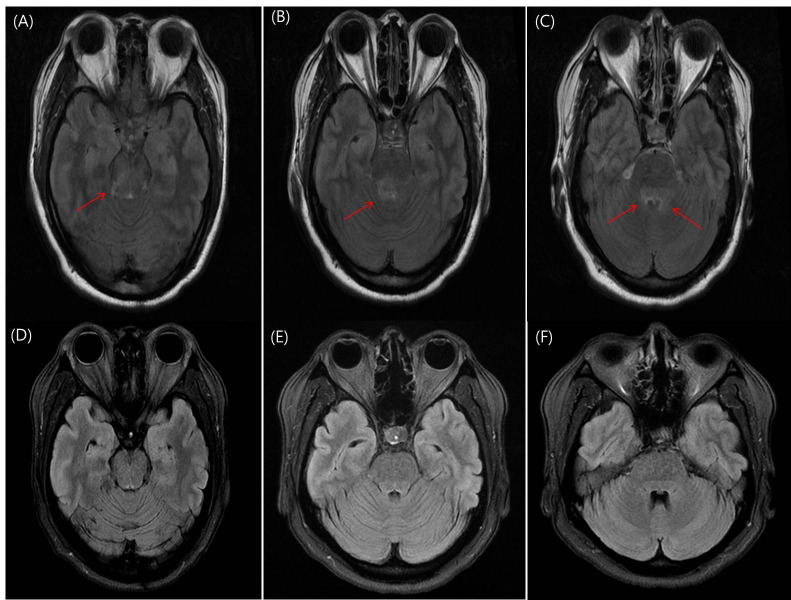
Abstract
Brainstem encephalitis (BE) associated with Mycoplasma pneumoniae in adults is rare, and the diagnosis is challenging. We describe an uncommon case of BE in an immunocompetent patient. A 43-year-old, otherwise healthy woman visited our emergency department with high fever and a sore throat, and 3 days later she returned with an altered drowsy mental status. Magnetic resonance imaging displayed diffuse swelling in bilateral cerebral regions involving the bilateral pons. The sera tested positive for the immunoglobulin (Ig) M antibody against M. Pneumoniae as detected by an enzyme immunoassay (EIA), and on hospital day 10, the level of IgM index against M. pneumoniae further increased from 1.5 to 2.1. We changed the antibiotic regimen from vancomycin and ceftriaxone to clarithromycin based on detection of M. pneumoniae, and we added intravenous immunoglobulin. After one month, the patient fully recovered from the neurological deficits. A follow-up brain magnetic resonance imaging was performed, which showed completely resolved lesions. Particle agglutination assay (PA) and EIA are both largely used to diagnose M. pneumoniae. Compared to the PA test, the EIA test could be a reliable tool because it separately measures IgM and IgG antibodies. We diagnosed BE associated with M. pneumoniae through EIA with an increasing level of IgM in the acute and subacute paired sera. Early treatment with macrolide antibiotics resulted in a good outcome.

 求助内容:
求助内容: 应助结果提醒方式:


