A Rare Presentation of Multifocal Anaplastic Oligodendroglioma.

Q3 Medicine
Ravikanth Reddy
{"title":"A Rare Presentation of Multifocal Anaplastic Oligodendroglioma.","authors":"Ravikanth Reddy","doi":"10.4103/jmau.jmau_126_20","DOIUrl":null,"url":null,"abstract":"<p><p>Multifocal tumors are usually reported within the same cerebral hemisphere due to widespread dissemination along the white matter tracts. This case report describes the magnetic resonance imaging appearances of multifocal anaplastic oligodendroglioma in a 28-year-old adult male that showed three discrete heterogeneously enhancing cortical-based lesions in the left frontoparietal lobes. Left frontal craniotomy was performed and biopsy of the lesion was obtained, histopathology of which showed features of anaplastic oligodendroglioma.</p>","PeriodicalId":16340,"journal":{"name":"Journal of Microscopy and Ultrastructure","volume":"11 2","pages":"115-117"},"PeriodicalIF":0.0000,"publicationDate":"2023-04-01","publicationTypes":"Journal Article","fieldsOfStudy":null,"isOpenAccess":false,"openAccessPdf":"https://ftp.ncbi.nlm.nih.gov/pub/pmc/oa_pdf/e2/60/JMAU-11-115.PMC10337670.pdf","citationCount":"0","resultStr":null,"platform":"Semanticscholar","paperid":null,"PeriodicalName":"Journal of Microscopy and Ultrastructure","FirstCategoryId":"1085","ListUrlMain":"https://doi.org/10.4103/jmau.jmau_126_20","RegionNum":0,"RegionCategory":null,"ArticlePicture":[],"TitleCN":null,"AbstractTextCN":null,"PMCID":null,"EPubDate":"","PubModel":"","JCR":"Q3","JCRName":"Medicine","Score":null,"Total":0}
引用次数: 0

Abstract

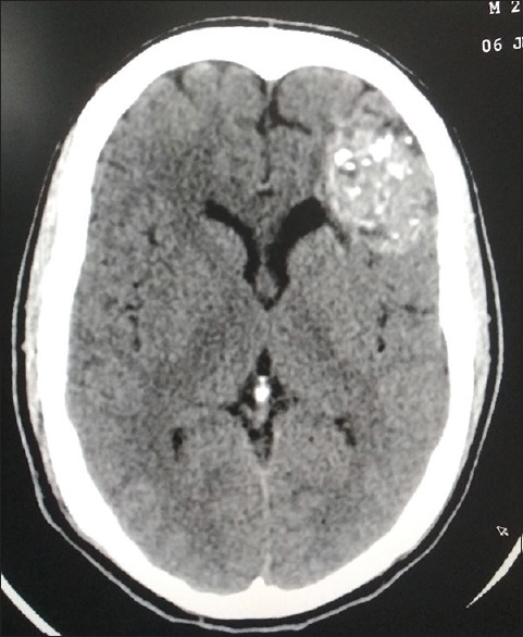
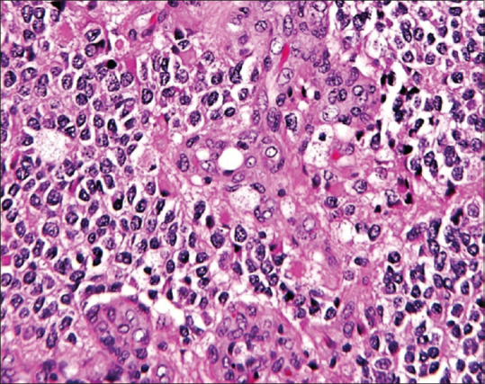
Multifocal tumors are usually reported within the same cerebral hemisphere due to widespread dissemination along the white matter tracts. This case report describes the magnetic resonance imaging appearances of multifocal anaplastic oligodendroglioma in a 28-year-old adult male that showed three discrete heterogeneously enhancing cortical-based lesions in the left frontoparietal lobes. Left frontal craniotomy was performed and biopsy of the lesion was obtained, histopathology of which showed features of anaplastic oligodendroglioma.
Abstract Image

Abstract Image

Abstract Image

一例罕见的多灶性间变性少突胶质细胞瘤。
由于沿白质束广泛播散,多灶性肿瘤通常在同一大脑半球内报道。本病例报告描述了一名28岁成年男性的多灶性间变性少突胶质细胞瘤的磁共振成像表现,在左侧额顶叶显示三个离散的异质强化皮层病变。左额叶开颅活检,病理表现为间变性少突胶质细胞瘤。
本文章由计算机程序翻译,如有差异,请以英文原文为准。
求助全文
约1分钟内获得全文 求助全文
来源期刊
CiteScore
1.90
自引率
0.00%
发文量
46
×
引用
GB/T 7714-2015
复制
MLA
复制
APA
复制
导出至
BibTeX EndNote RefMan NoteFirst NoteExpress
×
提示
您的信息不完整,为了账户安全,请先补充。
现在去补充
×
提示
您因"违规操作"
具体请查看互助需知
我知道了
×
提示
确定
请完成安全验证×
copy
已复制链接
快去分享给好友吧!
我知道了
右上角分享
点击右上角分享
0
联系我们:info@booksci.cn Book学术提供免费学术资源搜索服务,方便国内外学者检索中英文文献。致力于提供最便捷和优质的服务体验。 Copyright © 2023 布克学术 All rights reserved.
京ICP备2023020795号-1
ghs 京公网安备 11010802042870号
Book学术文献互助
Book学术文献互助群
群 号:481959085
Book学术官方微信