Simultaneous exercise stress cardiac magnetic resonance and cardiopulmonary exercise testing to elucidate the Fick components of aerobic exercise capacity: a feasibility and reproducibility study and pilot study in hematologic cancer survivors.
Justin M Canada, John McCarty, Jennifer H Jordan, Cory R Trankle, Kevin DeCamp, Josh D West, Mary Ann Reynolds, Rachel Myers, Katey Sweat, Virginia McGhee, Ross Arena, Antonio Abbate, W Gregory Hundley
{"title":"Simultaneous exercise stress cardiac magnetic resonance and cardiopulmonary exercise testing to elucidate the Fick components of aerobic exercise capacity: a feasibility and reproducibility study and pilot study in hematologic cancer survivors.","authors":"Justin M Canada, John McCarty, Jennifer H Jordan, Cory R Trankle, Kevin DeCamp, Josh D West, Mary Ann Reynolds, Rachel Myers, Katey Sweat, Virginia McGhee, Ross Arena, Antonio Abbate, W Gregory Hundley","doi":"10.1186/s40959-023-00182-1","DOIUrl":null,"url":null,"abstract":"<p><strong>Background: </strong>Patients treated for hematologic malignancy often experience reduced exercise capacity and increased fatigue; however whether this reduction is related to cardiac dysfunction or impairment of skeletal muscle oxygen extraction during activity is unknown. Cardiopulmonary exercise testing (CPET) coupled with stress cardiac magnetic resonance (ExeCMR), may provide a noninvasive method to identify the abnormalities of cardiac function or skeletal muscle oxygen extraction. This study was performed to determine the feasibility and reproducibility of a ExeCMR + CPET technique to measure the Fick components of peak oxygen consumption (VO<sub>2</sub>) and pilot its discriminatory potential in hematologic cancer patients experiencing fatigue.</p><p><strong>Methods: </strong>We studied 16 individuals undergoing ExeCMR to determine exercise cardiac reserve with simultaneous measures of VO<sub>2</sub>. The arteriovenous oxygen content difference (a-vO<sub>2</sub>diff) was calculated as the quotient of VO<sub>2</sub>/cardiac index (CI). Repeatability in measurements of peak VO<sub>2</sub>, CI, and a-vO<sub>2</sub>diff was assessed in seven healthy controls. Finally, we measured the Fick determinants of peak VO<sub>2</sub> in hematologic cancer survivors with fatigue (n = 6) and compared them to age/gender-matched healthy controls (n = 6).</p><p><strong>Results: </strong>Study procedures were successfully completed without any adverse events in all subjects (N = 16, 100%). The protocol demonstrated good-excellent test-retest reproducibility for peak VO<sub>2</sub> (intraclass correlation coefficient [ICC] = 0.992 [95%CI:0.955-0.999]; P < 0.001), peak CI (ICC = 0.970 [95%CI:0.838-0.995]; P < 0.001), and a-vO<sub>2</sub>diff (ICC = 0.953 [95%CI:0.744-0.992]; P < 0.001). Hematologic cancer survivors with fatigue demonstrated a significantly lower peak VO<sub>2</sub> (17.1 [13.5-23.5] vs. 26.0 [19.7-29.5] mL·kg<sup>-1</sup>·min<sup>-1</sup>, P = 0.026) and lower peak CI (5.0 [4.7-6.3] vs. 7.4 [7.0-8.8] L·min<sup>-1</sup>/m<sup>2</sup>, P = 0.004) without a significant difference in a-vO<sub>2</sub>diff (14.4 [11.8-16.9] vs. 13.6 [10.9-15.4] mLO<sub>2</sub>/dL, P = 0.589).</p><p><strong>Conclusions: </strong>Noninvasive measurement of peak VO<sub>2</sub> Fick determinants is feasible and reliable with an ExeCMR + CPET protocol in those treated for a hematologic malignancy and may offer insight into the mechanisms of exercise intolerance in those experiencing fatigue.</p>","PeriodicalId":9804,"journal":{"name":"Cardio-oncology","volume":null,"pages":null},"PeriodicalIF":3.2000,"publicationDate":"2023-07-10","publicationTypes":"Journal Article","fieldsOfStudy":null,"isOpenAccess":false,"openAccessPdf":"https://www.ncbi.nlm.nih.gov/pmc/articles/PMC10331991/pdf/","citationCount":"0","resultStr":null,"platform":"Semanticscholar","paperid":null,"PeriodicalName":"Cardio-oncology","FirstCategoryId":"1085","ListUrlMain":"https://doi.org/10.1186/s40959-023-00182-1","RegionNum":0,"RegionCategory":null,"ArticlePicture":[],"TitleCN":null,"AbstractTextCN":null,"PMCID":null,"EPubDate":"","PubModel":"","JCR":"Q2","JCRName":"CARDIAC & CARDIOVASCULAR SYSTEMS","Score":null,"Total":0}
引用次数: 0
Abstract
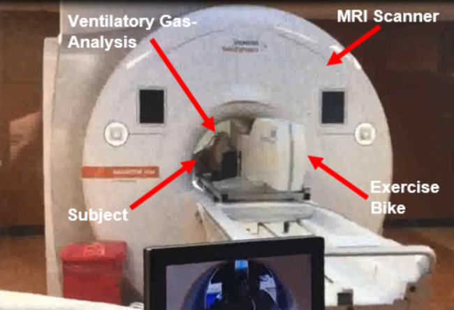
Background: Patients treated for hematologic malignancy often experience reduced exercise capacity and increased fatigue; however whether this reduction is related to cardiac dysfunction or impairment of skeletal muscle oxygen extraction during activity is unknown. Cardiopulmonary exercise testing (CPET) coupled with stress cardiac magnetic resonance (ExeCMR), may provide a noninvasive method to identify the abnormalities of cardiac function or skeletal muscle oxygen extraction. This study was performed to determine the feasibility and reproducibility of a ExeCMR + CPET technique to measure the Fick components of peak oxygen consumption (VO2) and pilot its discriminatory potential in hematologic cancer patients experiencing fatigue.
Methods: We studied 16 individuals undergoing ExeCMR to determine exercise cardiac reserve with simultaneous measures of VO2. The arteriovenous oxygen content difference (a-vO2diff) was calculated as the quotient of VO2/cardiac index (CI). Repeatability in measurements of peak VO2, CI, and a-vO2diff was assessed in seven healthy controls. Finally, we measured the Fick determinants of peak VO2 in hematologic cancer survivors with fatigue (n = 6) and compared them to age/gender-matched healthy controls (n = 6).
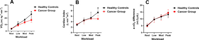
Results: Study procedures were successfully completed without any adverse events in all subjects (N = 16, 100%). The protocol demonstrated good-excellent test-retest reproducibility for peak VO2 (intraclass correlation coefficient [ICC] = 0.992 [95%CI:0.955-0.999]; P < 0.001), peak CI (ICC = 0.970 [95%CI:0.838-0.995]; P < 0.001), and a-vO2diff (ICC = 0.953 [95%CI:0.744-0.992]; P < 0.001). Hematologic cancer survivors with fatigue demonstrated a significantly lower peak VO2 (17.1 [13.5-23.5] vs. 26.0 [19.7-29.5] mL·kg-1·min-1, P = 0.026) and lower peak CI (5.0 [4.7-6.3] vs. 7.4 [7.0-8.8] L·min-1/m2, P = 0.004) without a significant difference in a-vO2diff (14.4 [11.8-16.9] vs. 13.6 [10.9-15.4] mLO2/dL, P = 0.589).
Conclusions: Noninvasive measurement of peak VO2 Fick determinants is feasible and reliable with an ExeCMR + CPET protocol in those treated for a hematologic malignancy and may offer insight into the mechanisms of exercise intolerance in those experiencing fatigue.
 求助内容:
求助内容: 应助结果提醒方式:


