Lena Mungenast , Ronya Nieminen , Carine Gaiser , Ana Bela Faia-Torres , Jürgen Rühe , Laura Suter-Dick
{"title":"Electrospun decellularized extracellular matrix scaffolds promote the regeneration of injured neurons","authors":"Lena Mungenast , Ronya Nieminen , Carine Gaiser , Ana Bela Faia-Torres , Jürgen Rühe , Laura Suter-Dick","doi":"10.1016/j.bbiosy.2023.100081","DOIUrl":null,"url":null,"abstract":"<div><p>Traumatic injury to the spinal cord (SCI) causes the transection of neurons, formation of a lesion cavity, and remodeling of the microenvironment by excessive extracellular matrix (ECM) deposition and scar formation leading to a regeneration-prohibiting environment. Electrospun fiber scaffolds have been shown to simulate the ECM and increase neural alignment and neurite outgrowth contributing to a growth-permissive matrix. In this work, electrospun ECM-like fibers providing biochemical and topological cues are implemented into a scaffold to represent an oriented biomaterial suitable for the alignment and migration of neural cells in order to improve spinal cord regeneration. The successfully decellularized spinal cord ECM (dECM), with no visible cell nuclei and dsDNA content < 50 ng/mg tissue, showed preserved ECM components, such as glycosaminoglycans and collagens. Serving as the biomaterial for 3D printer-assisted electrospinning, highly aligned and randomly distributed dECM fiber scaffolds (< 1 µm fiber diameter) were fabricated. The scaffolds were cytocompatible and supported the viability of a human neural cell line (SH-SY5Y) for 14 days. Cells were selectively differentiated into neurons, as confirmed by immunolabeling of specific cell markers (ChAT, Tubulin ß), and followed the orientation given by the dECM scaffolds. After generating a lesion site on the cell-scaffold model, cell migration was observed and compared to reference poly-ε-caprolactone fiber scaffolds. The aligned dECM fiber scaffold promoted the fastest and most efficient lesion closure, indicating superior cell guiding capabilities of dECM-based scaffolds. The strategy of combining decellularized tissues with controlled deposition of fibers to optimize biochemical and topographical cues opens the way for clinically relevant central nervous system scaffolding solutions.</p></div>","PeriodicalId":72379,"journal":{"name":"Biomaterials and biosystems","volume":"11 ","pages":"Article 100081"},"PeriodicalIF":0.0000,"publicationDate":"2023-09-01","publicationTypes":"Journal Article","fieldsOfStudy":null,"isOpenAccess":false,"openAccessPdf":"https://ftp.ncbi.nlm.nih.gov/pub/pmc/oa_pdf/9e/8e/main.PMC10329103.pdf","citationCount":"2","resultStr":null,"platform":"Semanticscholar","paperid":null,"PeriodicalName":"Biomaterials and biosystems","FirstCategoryId":"1085","ListUrlMain":"https://www.sciencedirect.com/science/article/pii/S2666534423000107","RegionNum":0,"RegionCategory":null,"ArticlePicture":[],"TitleCN":null,"AbstractTextCN":null,"PMCID":null,"EPubDate":"","PubModel":"","JCR":"Q3","JCRName":"Biochemistry, Genetics and Molecular Biology","Score":null,"Total":0}
引用次数: 2
Abstract
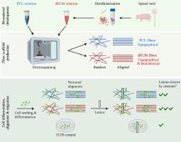
Traumatic injury to the spinal cord (SCI) causes the transection of neurons, formation of a lesion cavity, and remodeling of the microenvironment by excessive extracellular matrix (ECM) deposition and scar formation leading to a regeneration-prohibiting environment. Electrospun fiber scaffolds have been shown to simulate the ECM and increase neural alignment and neurite outgrowth contributing to a growth-permissive matrix. In this work, electrospun ECM-like fibers providing biochemical and topological cues are implemented into a scaffold to represent an oriented biomaterial suitable for the alignment and migration of neural cells in order to improve spinal cord regeneration. The successfully decellularized spinal cord ECM (dECM), with no visible cell nuclei and dsDNA content < 50 ng/mg tissue, showed preserved ECM components, such as glycosaminoglycans and collagens. Serving as the biomaterial for 3D printer-assisted electrospinning, highly aligned and randomly distributed dECM fiber scaffolds (< 1 µm fiber diameter) were fabricated. The scaffolds were cytocompatible and supported the viability of a human neural cell line (SH-SY5Y) for 14 days. Cells were selectively differentiated into neurons, as confirmed by immunolabeling of specific cell markers (ChAT, Tubulin ß), and followed the orientation given by the dECM scaffolds. After generating a lesion site on the cell-scaffold model, cell migration was observed and compared to reference poly-ε-caprolactone fiber scaffolds. The aligned dECM fiber scaffold promoted the fastest and most efficient lesion closure, indicating superior cell guiding capabilities of dECM-based scaffolds. The strategy of combining decellularized tissues with controlled deposition of fibers to optimize biochemical and topographical cues opens the way for clinically relevant central nervous system scaffolding solutions.

 求助内容:
求助内容: 应助结果提醒方式:


