Ectopic Cecal Varices as a Cause of Lower Gastrointestinal Bleeding.
IF 0.5
Q4 GASTROENTEROLOGY & HEPATOLOGY
引用次数: 0
Abstract
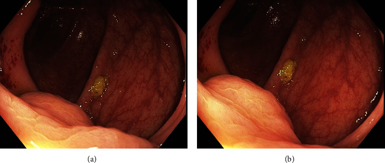
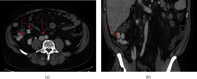
Ectopic varices account for 1%-5% of all variceal bleeding episodes in patients with portal hypertension. They can be found at any part of gastrointestinal tract including the small intestines, colon, or rectum. We report a case of a 59-year-old man who presented with bleeding per rectum 2 days after a routine colonoscopy, in which 2 lesions were biopsied. Gastroscopy was negative for bleeding, and he was not stable enough to undergo colonoscopy. CT angiography showed a large portosystemic shunt with multiple collaterals in the right lower quadrant. These findings were clues for a diagnosis of ectopic cecal varices.


盲肠异位静脉曲张是下消化道出血的原因之一。
异位静脉曲张占门脉高压患者所有静脉曲张出血发作的1%-5%。它们可以在胃肠道的任何部位发现,包括小肠、结肠或直肠。我们报告一个59岁的男性病例,他在常规结肠镜检查后2天出现直肠出血,其中2个病变进行了活检。胃镜检查未见出血,病情不稳定,不能进行结肠镜检查。CT血管造影显示右下象限一个大的门静脉系统分流伴多条侧枝。这些发现是诊断异位盲肠静脉曲张的线索。
本文章由计算机程序翻译,如有差异,请以英文原文为准。
求助全文
约1分钟内获得全文
求助全文
来源期刊

Case Reports in Gastrointestinal Medicine
GASTROENTEROLOGY & HEPATOLOGY-
自引率
0.00%
发文量
33
审稿时长
14 weeks
 求助内容:
求助内容: 应助结果提醒方式:


