Logan G Kirkland, Chloe G Garbe, Joseph Hadaya, Paul V Benson, Brant M Wagener, Sanjin Tankovic, Donald B Hoover
{"title":"Sympathetic innervation of human and porcine spleens: implications for between species variation in function.","authors":"Logan G Kirkland, Chloe G Garbe, Joseph Hadaya, Paul V Benson, Brant M Wagener, Sanjin Tankovic, Donald B Hoover","doi":"10.1186/s42234-022-00102-1","DOIUrl":null,"url":null,"abstract":"<p><strong>Background: </strong>The vagus nerve affects innate immune responses by activating spleen-projecting sympathetic neurons, which modulate leukocyte function. Recent basic and clinical research investigating vagus nerve stimulation to engage the cholinergic anti-inflammatory pathway (CAP) has shown promising therapeutic results for a variety of inflammatory diseases. Abundant sympathetic innervation occurs in rodent spleens, and use of these species has dominated mechanistic research investigating the CAP. However, previous neuroanatomical studies of human spleen found a more restricted pattern of innervation compared to rodents. Therefore, our primary goal was to establish the full extent of sympathetic innervation of human spleens using donor tissue with the shortest procurement to fixation time. Parallel studies of porcine spleen, a large animal model, were performed as a positive control and for comparison.</p><p><strong>Methods: </strong>Human and porcine spleen tissue were fixed immediately after harvest and prepared for immunohistochemistry. Human heart and porcine spleen were stained in conjunction as positive controls. Several immunohistochemical protocols were compared for best results. Tissue was stained for tyrosine hydroxylase (TH), a noradrenergic marker, using VIP purple chromogen. Consecutive tissue slices were stained for neuropeptide Y (NPY), which often co-localizes with TH, or double-labelled for TH and CD3, a T cell marker. High-magnification images and full scans of the tissue were obtained and analyzed for qualitative differences between species.</p><p><strong>Results: </strong>TH had dominant perivascular localization in human spleen, with negligible innervation of parenchyma, but such nerves were abundant throughout ventricular myocardium. In marked contrast, noradrenergic innervation was abundant in all regions of porcine spleen, with red pulp having more nerves than white pulp. NPY stain results were consistent with this pattern. In human spleen, noradrenergic nerves only ran close to T cells at the boundary of the periarterial lymphatic sheath and arteries. In porcine spleen, noradrenergic nerves were closely associated with T cells in both white and red pulp as well as other leukocytes in red pulp.</p><p><strong>Conclusion: </strong>Sympathetic innervation of the spleen varies between species in both distribution and abundance, with humans and pigs being at opposite extremes. This has important implications for sympathetic regulation of neuroimmune interactions in the spleen of different species and focused targeting of the CAP in humans.</p>","PeriodicalId":72363,"journal":{"name":"Bioelectronic medicine","volume":"8 1","pages":"20"},"PeriodicalIF":0.0000,"publicationDate":"2022-12-19","publicationTypes":"Journal Article","fieldsOfStudy":null,"isOpenAccess":false,"openAccessPdf":"https://www.ncbi.nlm.nih.gov/pmc/articles/PMC9762010/pdf/","citationCount":"2","resultStr":null,"platform":"Semanticscholar","paperid":null,"PeriodicalName":"Bioelectronic medicine","FirstCategoryId":"1085","ListUrlMain":"https://doi.org/10.1186/s42234-022-00102-1","RegionNum":0,"RegionCategory":null,"ArticlePicture":[],"TitleCN":null,"AbstractTextCN":null,"PMCID":null,"EPubDate":"","PubModel":"","JCR":"","JCRName":"","Score":null,"Total":0}
引用次数: 2
Abstract
Background: The vagus nerve affects innate immune responses by activating spleen-projecting sympathetic neurons, which modulate leukocyte function. Recent basic and clinical research investigating vagus nerve stimulation to engage the cholinergic anti-inflammatory pathway (CAP) has shown promising therapeutic results for a variety of inflammatory diseases. Abundant sympathetic innervation occurs in rodent spleens, and use of these species has dominated mechanistic research investigating the CAP. However, previous neuroanatomical studies of human spleen found a more restricted pattern of innervation compared to rodents. Therefore, our primary goal was to establish the full extent of sympathetic innervation of human spleens using donor tissue with the shortest procurement to fixation time. Parallel studies of porcine spleen, a large animal model, were performed as a positive control and for comparison.
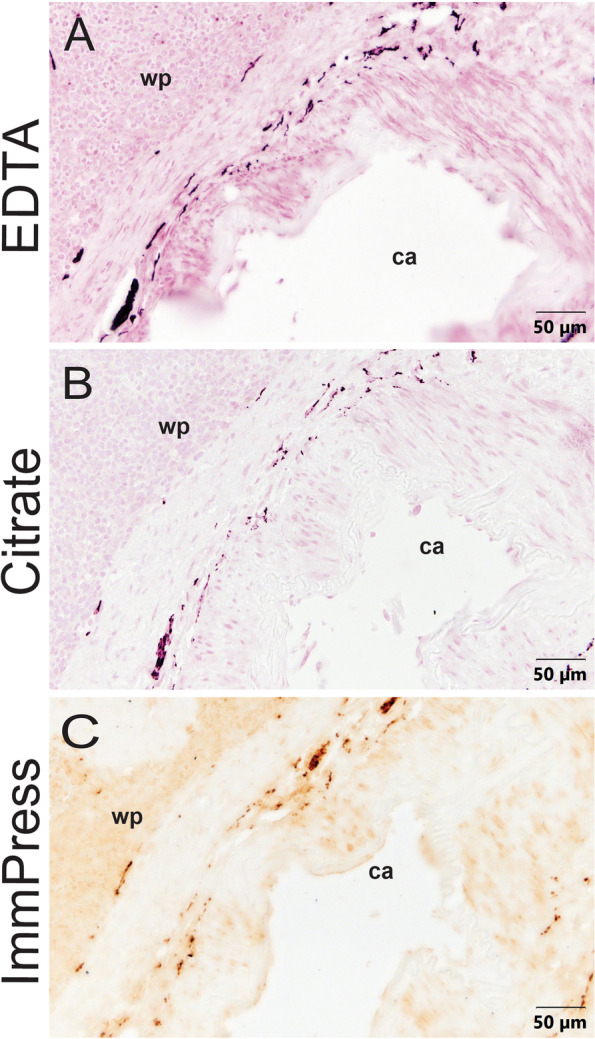
Methods: Human and porcine spleen tissue were fixed immediately after harvest and prepared for immunohistochemistry. Human heart and porcine spleen were stained in conjunction as positive controls. Several immunohistochemical protocols were compared for best results. Tissue was stained for tyrosine hydroxylase (TH), a noradrenergic marker, using VIP purple chromogen. Consecutive tissue slices were stained for neuropeptide Y (NPY), which often co-localizes with TH, or double-labelled for TH and CD3, a T cell marker. High-magnification images and full scans of the tissue were obtained and analyzed for qualitative differences between species.
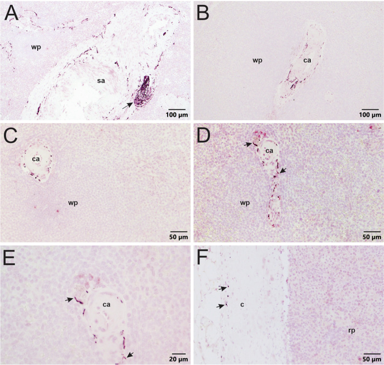
Results: TH had dominant perivascular localization in human spleen, with negligible innervation of parenchyma, but such nerves were abundant throughout ventricular myocardium. In marked contrast, noradrenergic innervation was abundant in all regions of porcine spleen, with red pulp having more nerves than white pulp. NPY stain results were consistent with this pattern. In human spleen, noradrenergic nerves only ran close to T cells at the boundary of the periarterial lymphatic sheath and arteries. In porcine spleen, noradrenergic nerves were closely associated with T cells in both white and red pulp as well as other leukocytes in red pulp.
Conclusion: Sympathetic innervation of the spleen varies between species in both distribution and abundance, with humans and pigs being at opposite extremes. This has important implications for sympathetic regulation of neuroimmune interactions in the spleen of different species and focused targeting of the CAP in humans.

 求助内容:
求助内容: 应助结果提醒方式:


