Wenxiu Zhang, Ying Duan, Lei Qi, Zhimei Li, Jiechuan Ren, Naluyele Nangale, Chunlan Yang
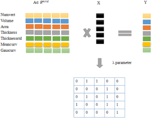
{"title":"Distinguishing Patients with MRI-Negative Temporal Lobe Epilepsy from Normal Controls Based on Individual Morphological Brain Network.","authors":"Wenxiu Zhang, Ying Duan, Lei Qi, Zhimei Li, Jiechuan Ren, Naluyele Nangale, Chunlan Yang","doi":"10.1007/s10548-023-00962-z","DOIUrl":null,"url":null,"abstract":"<p><p>Temporal Lobe Epilepsy (TLE) is the most common subtype of focal epilepsy and the most refractory to drug treatment. Roughly 30% of patients do not have easily identifiable structural abnormalities. In other words, MRI-negative TLE has normal MRI scans on visual inspection. Thus, MRI-negative TLE is a diagnostic and therapeutic challenge. In this study, we investigate the cortical morphological brain network to identify MRI-negative TLE. The 210 cortical ROIs based on the Brainnetome atlas were used to define the network nodes. The least absolute shrinkage and selection operator (LASSO) algorithm and Pearson correlation methods were used to calculate the inter-regional morphometric features vector correlation respectively. As a result, two types of networks were constructed. The topological characteristics of networks were calculated by graph theory. Then after, a two-stage feature selection strategy, including a two-sample t-test and support vector machine-based recursive feature elimination (SVM-RFE), was performed in feature selection. Finally, classification with support vector machine (SVM) and leave-one-out cross-validation (LOOCV) was employed for the training and evaluation of the classifiers. The performance of two constructed brain networks was compared in MRI-negative TLE classification. The results indicated that the LASSO algorithm achieved better performance than the Pearson pairwise correlation method. The LASSO algorithm provides a robust method of individual morphological network construction for distinguishing patients with MRI-negative TLE from normal controls.</p>","PeriodicalId":55329,"journal":{"name":"Brain Topography","volume":"36 4","pages":"554-565"},"PeriodicalIF":2.3000,"publicationDate":"2023-07-01","publicationTypes":"Journal Article","fieldsOfStudy":null,"isOpenAccess":false,"openAccessPdf":"","citationCount":"0","resultStr":null,"platform":"Semanticscholar","paperid":null,"PeriodicalName":"Brain Topography","FirstCategoryId":"3","ListUrlMain":"https://doi.org/10.1007/s10548-023-00962-z","RegionNum":3,"RegionCategory":"医学","ArticlePicture":[],"TitleCN":null,"AbstractTextCN":null,"PMCID":null,"EPubDate":"","PubModel":"","JCR":"Q3","JCRName":"CLINICAL NEUROLOGY","Score":null,"Total":0}
引用次数: 0
Abstract
Temporal Lobe Epilepsy (TLE) is the most common subtype of focal epilepsy and the most refractory to drug treatment. Roughly 30% of patients do not have easily identifiable structural abnormalities. In other words, MRI-negative TLE has normal MRI scans on visual inspection. Thus, MRI-negative TLE is a diagnostic and therapeutic challenge. In this study, we investigate the cortical morphological brain network to identify MRI-negative TLE. The 210 cortical ROIs based on the Brainnetome atlas were used to define the network nodes. The least absolute shrinkage and selection operator (LASSO) algorithm and Pearson correlation methods were used to calculate the inter-regional morphometric features vector correlation respectively. As a result, two types of networks were constructed. The topological characteristics of networks were calculated by graph theory. Then after, a two-stage feature selection strategy, including a two-sample t-test and support vector machine-based recursive feature elimination (SVM-RFE), was performed in feature selection. Finally, classification with support vector machine (SVM) and leave-one-out cross-validation (LOOCV) was employed for the training and evaluation of the classifiers. The performance of two constructed brain networks was compared in MRI-negative TLE classification. The results indicated that the LASSO algorithm achieved better performance than the Pearson pairwise correlation method. The LASSO algorithm provides a robust method of individual morphological network construction for distinguishing patients with MRI-negative TLE from normal controls.
期刊介绍:
Brain Topography publishes clinical and basic research on cognitive neuroscience and functional neurophysiology using the full range of imaging techniques including EEG, MEG, fMRI, TMS, diffusion imaging, spectroscopy, intracranial recordings, lesion studies, and related methods. Submissions combining multiple techniques are particularly encouraged, as well as reports of new and innovative methodologies.
 求助内容:
求助内容: 应助结果提醒方式:


