Comparison of Marginal Bone Loss in Simultaneous Versus Delayed Implant Placement Following Horizontal Ridge Augmentation with Autogenous Lateral Ramus Bone Block.
{"title":"Comparison of Marginal Bone Loss in Simultaneous Versus Delayed Implant Placement Following Horizontal Ridge Augmentation with Autogenous Lateral Ramus Bone Block.","authors":"Reza Tabrizi, Hassan Mohajerani, Hamidreza Moslemi, Shervin Shafiei, Shobeir Majdi","doi":"10.30476/dentjods.2022.92419.1641","DOIUrl":null,"url":null,"abstract":"<p><strong>Statement of the problem: </strong>Alveolar ridge resorption after tooth extraction may interfere with optimal dental implant placement.</p><p><strong>Purpose: </strong>This study aimed to compare the marginal bone loss (MBL) and thickness of the buccal aspect of the augmented site in simultaneous versus delayed implant placement following lateral ramus horizontal ridge augmentation in the posterior mandible.</p><p><strong>Materials and method: </strong>This prospective cohort study was conducted on patients who required horizontal bone augmentation of the posterior mandible using lateral ramus autogenous bone graft. Patients were divided into two groups of simultaneous implant placement (group 1) and delayed implant placement (group 2). Cone-beam computed tomography (CBCT) images were obtained before augmentation, at the time of implant placement, and 10 months later (6 months after implant loading). MBL and thickness of the buccal aspect were evaluated over time.</p><p><strong>Results: </strong>There were 18 patients in the group 1 and 16 patients in the group 2. Analysis of the CBCT scans demonstrated that the mean MBL was 1.21±0.35mm in the group 1 and 1.08±0.19mm in the group 2, with no significant difference between the two groups (<i>p</i>= 0.19). Thickness of the buccal aspect of the augmented site at the time of implant placement was 1.85±0.20mm in the group 1 and 2.16±0.29 mm in the group 2, with a significant difference (<i>p</i>< 0.001). However, data analysis regarding changes in the buccal plate thickness showed no significant difference between the two groups (<i>p</i>= 0.36).</p><p><strong>Conclusion: </strong>According to the results of this study, there was no significant difference in M-BL and post-operative changes in the thickness of the buccal aspect of the augmented sites with onlay lateral ramus bone blocks between simultaneous and delayed implant placement.</p>","PeriodicalId":73702,"journal":{"name":"Journal of dentistry (Shiraz, Iran)","volume":"24 2","pages":"200-205"},"PeriodicalIF":0.0000,"publicationDate":"2023-06-01","publicationTypes":"Journal Article","fieldsOfStudy":null,"isOpenAccess":false,"openAccessPdf":"https://www.ncbi.nlm.nih.gov/pmc/articles/PMC10300137/pdf/","citationCount":"0","resultStr":null,"platform":"Semanticscholar","paperid":null,"PeriodicalName":"Journal of dentistry (Shiraz, Iran)","FirstCategoryId":"1085","ListUrlMain":"https://doi.org/10.30476/dentjods.2022.92419.1641","RegionNum":0,"RegionCategory":null,"ArticlePicture":[],"TitleCN":null,"AbstractTextCN":null,"PMCID":null,"EPubDate":"","PubModel":"","JCR":"","JCRName":"","Score":null,"Total":0}
引用次数: 0
Abstract
Statement of the problem: Alveolar ridge resorption after tooth extraction may interfere with optimal dental implant placement.
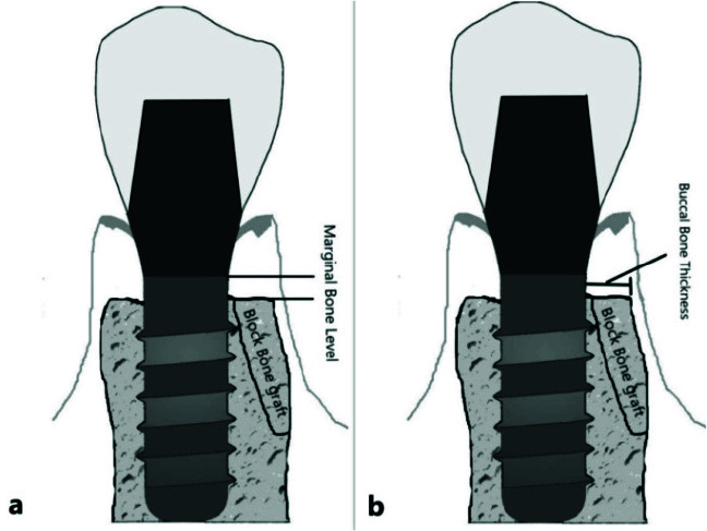
Purpose: This study aimed to compare the marginal bone loss (MBL) and thickness of the buccal aspect of the augmented site in simultaneous versus delayed implant placement following lateral ramus horizontal ridge augmentation in the posterior mandible.
Materials and method: This prospective cohort study was conducted on patients who required horizontal bone augmentation of the posterior mandible using lateral ramus autogenous bone graft. Patients were divided into two groups of simultaneous implant placement (group 1) and delayed implant placement (group 2). Cone-beam computed tomography (CBCT) images were obtained before augmentation, at the time of implant placement, and 10 months later (6 months after implant loading). MBL and thickness of the buccal aspect were evaluated over time.
Results: There were 18 patients in the group 1 and 16 patients in the group 2. Analysis of the CBCT scans demonstrated that the mean MBL was 1.21±0.35mm in the group 1 and 1.08±0.19mm in the group 2, with no significant difference between the two groups (p= 0.19). Thickness of the buccal aspect of the augmented site at the time of implant placement was 1.85±0.20mm in the group 1 and 2.16±0.29 mm in the group 2, with a significant difference (p< 0.001). However, data analysis regarding changes in the buccal plate thickness showed no significant difference between the two groups (p= 0.36).
Conclusion: According to the results of this study, there was no significant difference in M-BL and post-operative changes in the thickness of the buccal aspect of the augmented sites with onlay lateral ramus bone blocks between simultaneous and delayed implant placement.

 求助内容:
求助内容: 应助结果提醒方式:


