Jason Gan, Romman Nourzaie, Brent J Doolan, Steve Connor
{"title":"CT and MRI of head and neck cutaneous lesions.","authors":"Jason Gan, Romman Nourzaie, Brent J Doolan, Steve Connor","doi":"10.1259/bjro.20230006","DOIUrl":null,"url":null,"abstract":"<p><p>Cutaneous lesions are derived from the epidermis, dermis and cutaneous appendages. Whilst imaging may occasionally be performed to evaluate such lesions, they may be undiagnosed and demonstrated for the first time on head and neck imaging studies. Although usually amenable to clinical examination and biopsy, CT or MRI studies may also demonstrate characteristic imaging features which aid the radiological differential diagnosis. In addition, imaging studies define the extent and staging of malignant lesions, as well as the complications of benign lesions. It is important for the radiologist to understanding the clinical significance and associations of these cutaneous conditions. This pictorial review will describe and depict the imaging appearances of benign, malignant, overgrowth, blistering, appendage and syndromic cutaneous lesions. An increasing awareness of the imaging characteristics of cutaneous lesions and related conditions will help the framing of a clinically relevant report.</p>","PeriodicalId":72419,"journal":{"name":"BJR open","volume":"5 1","pages":"20230006"},"PeriodicalIF":2.1000,"publicationDate":"2023-01-01","publicationTypes":"Journal Article","fieldsOfStudy":null,"isOpenAccess":false,"openAccessPdf":"https://www.ncbi.nlm.nih.gov/pmc/articles/PMC10301713/pdf/","citationCount":"0","resultStr":null,"platform":"Semanticscholar","paperid":null,"PeriodicalName":"BJR open","FirstCategoryId":"1085","ListUrlMain":"https://doi.org/10.1259/bjro.20230006","RegionNum":0,"RegionCategory":null,"ArticlePicture":[],"TitleCN":null,"AbstractTextCN":null,"PMCID":null,"EPubDate":"","PubModel":"","JCR":"","JCRName":"","Score":null,"Total":0}
引用次数: 0
Abstract
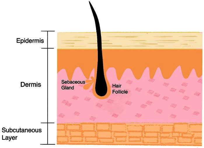
Cutaneous lesions are derived from the epidermis, dermis and cutaneous appendages. Whilst imaging may occasionally be performed to evaluate such lesions, they may be undiagnosed and demonstrated for the first time on head and neck imaging studies. Although usually amenable to clinical examination and biopsy, CT or MRI studies may also demonstrate characteristic imaging features which aid the radiological differential diagnosis. In addition, imaging studies define the extent and staging of malignant lesions, as well as the complications of benign lesions. It is important for the radiologist to understanding the clinical significance and associations of these cutaneous conditions. This pictorial review will describe and depict the imaging appearances of benign, malignant, overgrowth, blistering, appendage and syndromic cutaneous lesions. An increasing awareness of the imaging characteristics of cutaneous lesions and related conditions will help the framing of a clinically relevant report.

 求助内容:
求助内容: 应助结果提醒方式:


