Positive Rate of Malignant Cells in Endometrial Cytology Samples of Ovarian Cancer, Fallopian Tube Cancer, and Primary Peritoneal Cancer Patients: A Systematic Review and Meta-Analysis.
{"title":"Positive Rate of Malignant Cells in Endometrial Cytology Samples of Ovarian Cancer, Fallopian Tube Cancer, and Primary Peritoneal Cancer Patients: A Systematic Review and Meta-Analysis.","authors":"Tiantian Wang, Yadi Bin, Lanbo Zhao, Qiling Li","doi":"10.4103/joc.joc_49_22","DOIUrl":null,"url":null,"abstract":"<p><p>To estimate the feasibility of diagnosing ovarian cancer, fallopian tube cancer, and primary peritoneal cancer through endometrial cytology, we performed a systematic review and meta-analysis to calculate the pooled positive rate of malignant cells in endometrial cytology samples. We queried PubMed, EMBASE, Medline, and Cochrane Central Register of Controlled Trails from inception to November 12, 2020 for studies estimating positive rates of malignant cells in endometrial cytology samples from patients with ovarian cancer, fallopian tube cancer, and primary peritoneal cancer. The positive rates of the included studies were calculated as pooled positive rate through meta-analyses of proportion. Subgroup analysis based on different sampling methods was conducted. Seven retrospective studies involving 975 patients were included. Pooled positive rate of malignant cells in endometrial cytology specimens of ovarian cancer, fallopian tube cancer, and primary peritoneal cancer patients was 23% (95% CI: 16% - 34%). Statistical heterogeneity between the included studies was considerable (<i>I</i><sup>2</sup> = 89%, <i>P</i> < 0.01). The pooled positive rates of the group of brushes and the group of aspiration smears were 13% (95% CI: 10% - 17%, <i>I</i><sup>2</sup> = 0, <i>P</i> = 0.45) and 33% (95% CI: 25% - 42%, <i>I</i><sup>2</sup> = 80%, <i>P</i> < 0.01), respectively. Although endometrial cytology is not an ideal diagnostic tool for ovarian cancer, fallopian tube cancer, and primary peritoneal cancer, it is a convenient, painless, and easy-to-implement adjunct to other tools. Sampling method is one of the factors that affect the detection rate.</p>","PeriodicalId":50217,"journal":{"name":"Journal of Cytology","volume":"40 2","pages":"51-57"},"PeriodicalIF":1.0000,"publicationDate":"2023-04-01","publicationTypes":"Journal Article","fieldsOfStudy":null,"isOpenAccess":false,"openAccessPdf":"https://www.ncbi.nlm.nih.gov/pmc/articles/PMC10305903/pdf/","citationCount":"0","resultStr":null,"platform":"Semanticscholar","paperid":null,"PeriodicalName":"Journal of Cytology","FirstCategoryId":"3","ListUrlMain":"https://doi.org/10.4103/joc.joc_49_22","RegionNum":4,"RegionCategory":"医学","ArticlePicture":[],"TitleCN":null,"AbstractTextCN":null,"PMCID":null,"EPubDate":"2023/5/22 0:00:00","PubModel":"Epub","JCR":"Q4","JCRName":"MEDICAL LABORATORY TECHNOLOGY","Score":null,"Total":0}
引用次数: 0
Abstract
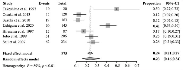
To estimate the feasibility of diagnosing ovarian cancer, fallopian tube cancer, and primary peritoneal cancer through endometrial cytology, we performed a systematic review and meta-analysis to calculate the pooled positive rate of malignant cells in endometrial cytology samples. We queried PubMed, EMBASE, Medline, and Cochrane Central Register of Controlled Trails from inception to November 12, 2020 for studies estimating positive rates of malignant cells in endometrial cytology samples from patients with ovarian cancer, fallopian tube cancer, and primary peritoneal cancer. The positive rates of the included studies were calculated as pooled positive rate through meta-analyses of proportion. Subgroup analysis based on different sampling methods was conducted. Seven retrospective studies involving 975 patients were included. Pooled positive rate of malignant cells in endometrial cytology specimens of ovarian cancer, fallopian tube cancer, and primary peritoneal cancer patients was 23% (95% CI: 16% - 34%). Statistical heterogeneity between the included studies was considerable (I2 = 89%, P < 0.01). The pooled positive rates of the group of brushes and the group of aspiration smears were 13% (95% CI: 10% - 17%, I2 = 0, P = 0.45) and 33% (95% CI: 25% - 42%, I2 = 80%, P < 0.01), respectively. Although endometrial cytology is not an ideal diagnostic tool for ovarian cancer, fallopian tube cancer, and primary peritoneal cancer, it is a convenient, painless, and easy-to-implement adjunct to other tools. Sampling method is one of the factors that affect the detection rate.
为了评估通过子宫内膜细胞学诊断卵巢癌症、癌症和原发性腹膜癌症的可行性,我们进行了系统回顾和荟萃分析,以计算子宫内膜细胞学样本中恶性细胞的合并阳性率。从开始到2020年11月12日,我们查询了PubMed、EMBASE、Medline和Cochrane Central Register of Controlled Trails,以了解估计卵巢癌症、癌症输卵管和原发性癌症患者子宫内膜细胞学样本中恶性细胞阳性率的研究。纳入研究的阳性率通过比例荟萃分析计算为合并阳性率。根据不同的抽样方法进行亚组分析。包括7项涉及975名患者的回顾性研究。卵巢癌症、癌症和原发性癌症患者子宫内膜细胞学标本中恶性细胞的合并阳性率为23%(95%CI:16%-34%)。纳入研究之间的统计学异质性相当大(I2=89%,P<0.01)。刷子组和抽吸涂片组的合并阳性率分别为13%(95%CI:10%-17%,I2=0,P=0.45)和33%(95%CI:25%-42%,I2=80%,P<0.01)。尽管子宫内膜细胞学检查不是卵巢癌症、癌症输卵管和原发性癌症的理想诊断工具,但它是其他工具的一种方便、无痛和易于实现的辅助工具。采样方法是影响检测率的因素之一。
期刊介绍:
The Journal of Cytology is the official Quarterly publication of the Indian Academy of Cytologists. It is in the 25th year of publication in the year 2008. The journal covers all aspects of diagnostic cytology, including fine needle aspiration cytology, gynecological and non-gynecological cytology. Articles on ancillary techniques, like cytochemistry, immunocytochemistry, electron microscopy, molecular cytopathology, as applied to cytological material are also welcome. The journal gives preference to clinically oriented studies over experimental and animal studies. The Journal would publish peer-reviewed original research papers, case reports, systematic reviews, meta-analysis, and debates.

 求助内容:
求助内容: 应助结果提醒方式:


