Enhanced Extraction of Blood and Tissue Time-Activity Curves in Cardiac Mouse FDG PET Imaging by Means of Constrained Nonnegative Matrix Factorization.
Otman Sarrhini, Pedro D'Orléans-Juste, Jacques A Rousseau, Jean-François Beaudoin, Roger Lecomte
{"title":"Enhanced Extraction of Blood and Tissue Time-Activity Curves in Cardiac Mouse FDG PET Imaging by Means of Constrained Nonnegative Matrix Factorization.","authors":"Otman Sarrhini, Pedro D'Orléans-Juste, Jacques A Rousseau, Jean-François Beaudoin, Roger Lecomte","doi":"10.1155/2023/5366733","DOIUrl":null,"url":null,"abstract":"<p><p>We propose an enhanced method to accurately retrieve time-activity curves (TACs) of blood and tissue from dynamic 2-deoxy-2-[<sup>18</sup>F]fluoro-D-glucose ([<sup>18</sup>F]FDG) positron emission tomography (PET) cardiac images of mice. The method is noninvasive and consists of using a constrained nonnegative matrix factorization algorithm (CNMF) applied to the matrix (<i>A</i>) containing the intensity values of the voxels of the left ventricle (LV) PET image. CNMF factorizes <i>A</i> into nonnegative matrices <i>H</i> and <i>W</i>, respectively, representing the physiological factors (blood and tissue) and their associated weights, by minimizing an extended cost function. We verified our method on 32 C57BL/6 mice, 14 of them with acute myocardial infarction (AMI). With CNMF, we could break down the mouse LV into myocardial and blood pool images. Their corresponding TACs were used in kinetic modeling to readily determine the [<sup>18</sup>F]FDG influx constant (<i>K</i><sub><i>i</i></sub>) required to compute the myocardial metabolic rate of glucose. The calculated <i>K</i><sub><i>i</i></sub> values using CNMF for the heart of control mice were in good agreement with those published in the literature. Significant differences in <i>K</i><sub><i>i</i></sub> values for the heart of control and AMI mice were found using CNMF. The values of the elements of <i>W</i> agreed well with the LV structural changes induced by ligation of the left coronary artery. CNMF was compared with the recently published method based on robust unmixing of dynamic sequences using regions of interest (RUDUR). A clear improvement of signal separation was observed with CNMF compared to the RUDUR method.</p>","PeriodicalId":47063,"journal":{"name":"International Journal of Biomedical Imaging","volume":"2023 ","pages":"5366733"},"PeriodicalIF":3.3000,"publicationDate":"2023-01-01","publicationTypes":"Journal Article","fieldsOfStudy":null,"isOpenAccess":false,"openAccessPdf":"https://www.ncbi.nlm.nih.gov/pmc/articles/PMC10287520/pdf/","citationCount":"2","resultStr":null,"platform":"Semanticscholar","paperid":null,"PeriodicalName":"International Journal of Biomedical Imaging","FirstCategoryId":"1085","ListUrlMain":"https://doi.org/10.1155/2023/5366733","RegionNum":0,"RegionCategory":null,"ArticlePicture":[],"TitleCN":null,"AbstractTextCN":null,"PMCID":null,"EPubDate":"","PubModel":"","JCR":"Q2","JCRName":"ENGINEERING, BIOMEDICAL","Score":null,"Total":0}
引用次数: 2
Abstract
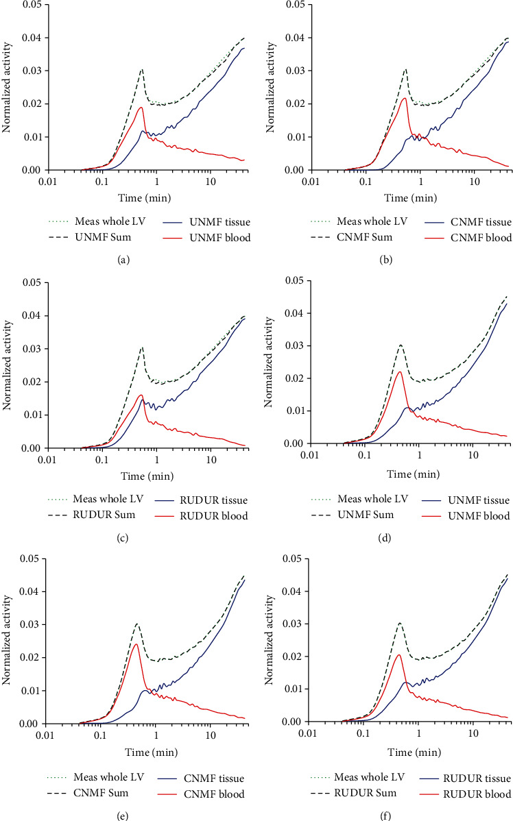
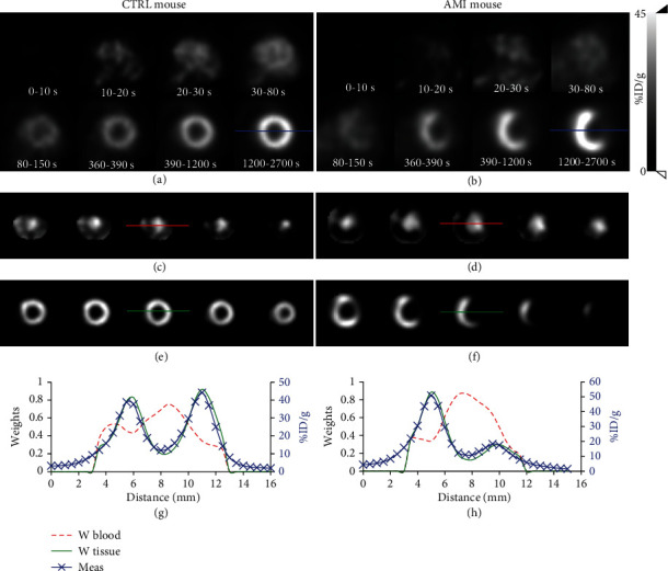
We propose an enhanced method to accurately retrieve time-activity curves (TACs) of blood and tissue from dynamic 2-deoxy-2-[18F]fluoro-D-glucose ([18F]FDG) positron emission tomography (PET) cardiac images of mice. The method is noninvasive and consists of using a constrained nonnegative matrix factorization algorithm (CNMF) applied to the matrix (A) containing the intensity values of the voxels of the left ventricle (LV) PET image. CNMF factorizes A into nonnegative matrices H and W, respectively, representing the physiological factors (blood and tissue) and their associated weights, by minimizing an extended cost function. We verified our method on 32 C57BL/6 mice, 14 of them with acute myocardial infarction (AMI). With CNMF, we could break down the mouse LV into myocardial and blood pool images. Their corresponding TACs were used in kinetic modeling to readily determine the [18F]FDG influx constant (Ki) required to compute the myocardial metabolic rate of glucose. The calculated Ki values using CNMF for the heart of control mice were in good agreement with those published in the literature. Significant differences in Ki values for the heart of control and AMI mice were found using CNMF. The values of the elements of W agreed well with the LV structural changes induced by ligation of the left coronary artery. CNMF was compared with the recently published method based on robust unmixing of dynamic sequences using regions of interest (RUDUR). A clear improvement of signal separation was observed with CNMF compared to the RUDUR method.
期刊介绍:
The International Journal of Biomedical Imaging is managed by a board of editors comprising internationally renowned active researchers. The journal is freely accessible online and also offered for purchase in print format. It employs a web-based review system to ensure swift turnaround times while maintaining high standards. In addition to regular issues, special issues are organized by guest editors. The subject areas covered include (but are not limited to):
Digital radiography and tomosynthesis
X-ray computed tomography (CT)
Magnetic resonance imaging (MRI)
Single photon emission computed tomography (SPECT)
Positron emission tomography (PET)
Ultrasound imaging
Diffuse optical tomography, coherence, fluorescence, bioluminescence tomography, impedance tomography
Neutron imaging for biomedical applications
Magnetic and optical spectroscopy, and optical biopsy
Optical, electron, scanning tunneling/atomic force microscopy
Small animal imaging
Functional, cellular, and molecular imaging
Imaging assays for screening and molecular analysis
Microarray image analysis and bioinformatics
Emerging biomedical imaging techniques
Imaging modality fusion
Biomedical imaging instrumentation
Biomedical image processing, pattern recognition, and analysis
Biomedical image visualization, compression, transmission, and storage
Imaging and modeling related to systems biology and systems biomedicine
Applied mathematics, applied physics, and chemistry related to biomedical imaging
Grid-enabling technology for biomedical imaging and informatics


 求助内容:
求助内容: 应助结果提醒方式:


