Brain Perfusion Changes in a Patient with Facial Trauma.

IF 0.9 Q4 RADIOLOGY, NUCLEAR MEDICINE & MEDICAL IMAGING
Chrissa Sioka, Anastasia Zikou, Petros Petrikis, Asimakis Asimakopoulos, George Alexiou, Vasileios Ragos
{"title":"Brain Perfusion Changes in a Patient with Facial Trauma.","authors":"Chrissa Sioka,&nbsp;Anastasia Zikou,&nbsp;Petros Petrikis,&nbsp;Asimakis Asimakopoulos,&nbsp;George Alexiou,&nbsp;Vasileios Ragos","doi":"10.4274/mirt.galenos.2022.90958","DOIUrl":null,"url":null,"abstract":"<p><p>A 69-year-old male was admitted to our hospital because of left facial trauma with bone fractures, including the maxillary sinus, zygomatic arch, and ethmoid and sphenoid bones. Brain computed tomography was unremarkable but regional cerebral blood flow with hexamethyl-propylene-amine oxime single-photon emission computed tomography (SPECT) showed hypoperfusion of the left hemisphere, which was reversible since a repeat SPECT 4 months later was substantially improved. Brain perfusion SPECT may provide information on cerebrovascular status in some cases of facial injury.</p>","PeriodicalId":44681,"journal":{"name":"Molecular Imaging and Radionuclide Therapy","volume":null,"pages":null},"PeriodicalIF":0.9000,"publicationDate":"2023-06-20","publicationTypes":"Journal Article","fieldsOfStudy":null,"isOpenAccess":false,"openAccessPdf":"https://ftp.ncbi.nlm.nih.gov/pub/pmc/oa_pdf/33/ff/MIRT-32-162.PMC10284177.pdf","citationCount":"0","resultStr":null,"platform":"Semanticscholar","paperid":null,"PeriodicalName":"Molecular Imaging and Radionuclide Therapy","FirstCategoryId":"1085","ListUrlMain":"https://doi.org/10.4274/mirt.galenos.2022.90958","RegionNum":0,"RegionCategory":null,"ArticlePicture":[],"TitleCN":null,"AbstractTextCN":null,"PMCID":null,"EPubDate":"","PubModel":"","JCR":"Q4","JCRName":"RADIOLOGY, NUCLEAR MEDICINE & MEDICAL IMAGING","Score":null,"Total":0}
引用次数: 0

Abstract

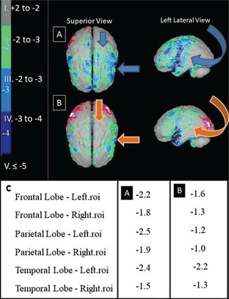
A 69-year-old male was admitted to our hospital because of left facial trauma with bone fractures, including the maxillary sinus, zygomatic arch, and ethmoid and sphenoid bones. Brain computed tomography was unremarkable but regional cerebral blood flow with hexamethyl-propylene-amine oxime single-photon emission computed tomography (SPECT) showed hypoperfusion of the left hemisphere, which was reversible since a repeat SPECT 4 months later was substantially improved. Brain perfusion SPECT may provide information on cerebrovascular status in some cases of facial injury.
Abstract Image

Abstract Image

面部创伤患者的脑灌注改变。
一名69岁男性因左侧面部外伤合并骨折,包括上颌窦、颧弓、筛骨和蝶骨入院。脑部计算机断层扫描无明显异常,但六甲基丙烯胺肟单光子发射计算机断层扫描(SPECT)显示左半球脑血流不足,由于4个月后重复SPECT明显改善,这是可逆的。脑灌注SPECT可以提供一些面部损伤病例的脑血管状态信息。
本文章由计算机程序翻译,如有差异,请以英文原文为准。
求助全文
约1分钟内获得全文 求助全文
来源期刊
Molecular Imaging and Radionuclide Therapy
Molecular Imaging and Radionuclide Therapy RADIOLOGY, NUCLEAR MEDICINE & MEDICAL IMAGING-
CiteScore
1.30
自引率
0.00%
发文量
50
期刊介绍: Molecular Imaging and Radionuclide Therapy (Mol Imaging Radionucl Ther, MIRT) is publishes original research articles, invited reviews, editorials, short communications, letters, consensus statements, guidelines and case reports with a literature review on the topic, in the field of molecular imaging, multimodality imaging, nuclear medicine, radionuclide therapy, radiopharmacy, medical physics, dosimetry and radiobiology.
×
引用
GB/T 7714-2015
复制
MLA
复制
APA
复制
导出至
BibTeX EndNote RefMan NoteFirst NoteExpress
×
提示
您的信息不完整,为了账户安全,请先补充。
现在去补充
×
提示
您因"违规操作"
具体请查看互助需知
我知道了
×
提示
确定
请完成安全验证×
copy
已复制链接
快去分享给好友吧!
我知道了
右上角分享
点击右上角分享
0
联系我们:info@booksci.cn Book学术提供免费学术资源搜索服务,方便国内外学者检索中英文文献。致力于提供最便捷和优质的服务体验。 Copyright © 2023 布克学术 All rights reserved.
京ICP备2023020795号-1
ghs 京公网安备 11010802042870号
Book学术文献互助
Book学术文献互助群
群 号:481959085
Book学术官方微信