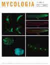
Using fimbrin to quantify the endocytic subapical collar during polarized growth in three filamentous fungi.
IF 2.6
2区 生物学
Q2 MYCOLOGY
引用次数: 0
Abstract
ABSTRACT Filamentous fungi produce specialized cells called hyphae. These cells grow by polarized extension at their apex, which is maintained by the balance of endocytosis and exocytosis at the apex. Although endocytosis has been well characterized in other organisms, the details of endocytosis and its role in maintaining polarity during hyphal growth in filamentous fungi is comparatively sparsely studied. In recent years, a concentrated region of protein activity that trails the growing apex of hyphal cells has been discovered. This region, dubbed the “endocytic collar” (EC), is a dynamic 3-dimensional region of concentrated endocytic activity, the disruption of which results in the loss of hyphal polarity. Here, fluorescent protein–tagged fimbrin was used as a marker to map the collar during growth of hyphae in three fungi: Aspergillus nidulans, Colletotrichum graminicola, and Neurospora crassa. Advanced microscopy techniques and novel quantification strategies were then utilized to quantify the spatiotemporal localization and recovery rates of fimbrin in the EC during hyphal growth. Correlating these variables with hyphal growth rate revealed that the strongest observed relationship with hyphal growth is the distance by which the EC trails the apex, and that measured endocytic rate does not correlate strongly with hyphal growth rate. This supports the hypothesis that endocytic influence on hyphal growth rate is better explained by spatiotemporal regulation of the EC than by the raw rate of endocytosis.利用纤维蛋白定量分析了三种丝状真菌极化生长过程中的内吞亚根尖环。
丝状真菌产生称为菌丝的特殊细胞。这些细胞在其顶端以极化伸展的方式生长,这是由顶端的内吞和胞吐平衡维持的。虽然内吞作用在其他生物中已经有很好的特征,但对丝状真菌中内吞作用的细节及其在菌丝生长过程中维持极性的作用的研究相对较少。近年来,在菌丝细胞的生长顶端发现了一个蛋白质活性集中的区域。这个区域被称为“内吞环”(EC),是一个动态的三维内吞活性集中区域,其破坏导致菌丝极性的丧失。本研究利用荧光蛋白标记的纤维蛋白作为标记物,绘制了三种真菌(灰曲霉、禾本科炭疽菌和粗神经孢子菌)菌丝生长过程中的领。然后利用先进的显微镜技术和新的量化策略来量化菌丝生长过程中纤维蛋白在EC中的时空定位和回收率。这些变量与菌丝生长速率的相关性表明,与菌丝生长最密切的关系是菌丝离顶的距离,而测定的内吞噬率与菌丝生长速率的相关性不强。这支持了一个假设,即内吞作用对菌丝生长速度的影响可以通过EC的时空调节来更好地解释,而不是通过内吞作用的原始速率。
本文章由计算机程序翻译,如有差异,请以英文原文为准。
求助全文
约1分钟内获得全文
求助全文
来源期刊

Mycologia
生物-真菌学
CiteScore
6.20
自引率
3.60%
发文量
56
审稿时长
4-8 weeks
期刊介绍:
International in coverage, Mycologia presents recent advances in mycology, emphasizing all aspects of the biology of Fungi and fungus-like organisms, including Lichens, Oomycetes and Slime Molds. The Journal emphasizes subjects including applied biology, biochemistry, cell biology, development, ecology, evolution, genetics, genomics, molecular biology, morphology, new techniques, animal or plant pathology, phylogenetics, physiology, aspects of secondary metabolism, systematics, and ultrastructure. In addition to research articles, reviews and short notes, Mycologia also includes invited papers based on presentations from the Annual Conference of the Mycological Society of America, such as Karling Lectures or Presidential Addresses.
 求助内容:
求助内容: 应助结果提醒方式:


