Clothilde Claus, Moriya Slavin, Eugénie Ansseau, Céline Lancelot, Karimatou Bah, Saskia Lassche, Manon Fiévet, Anna Greco, Sara Tomaiuolo, Alexandra Tassin, Virginie Dudome, Benno Kusters, Anne-Emilie Declèves, Dalila Laoudj-Chenivesse, Baziel G M van Engelen, Denis Nonclercq, Alexandra Belayew, Nir Kalisman, Frédérique Coppée
{"title":"The double homeodomain protein DUX4c is associated with regenerating muscle fibers and RNA-binding proteins.","authors":"Clothilde Claus, Moriya Slavin, Eugénie Ansseau, Céline Lancelot, Karimatou Bah, Saskia Lassche, Manon Fiévet, Anna Greco, Sara Tomaiuolo, Alexandra Tassin, Virginie Dudome, Benno Kusters, Anne-Emilie Declèves, Dalila Laoudj-Chenivesse, Baziel G M van Engelen, Denis Nonclercq, Alexandra Belayew, Nir Kalisman, Frédérique Coppée","doi":"10.1186/s13395-022-00310-y","DOIUrl":null,"url":null,"abstract":"<p><strong>Background: </strong>We have previously demonstrated that double homeobox 4 centromeric (DUX4C) encoded for a functional DUX4c protein upregulated in dystrophic skeletal muscles. Based on gain- and loss-of-function studies we have proposed DUX4c involvement in muscle regeneration. Here, we provide further evidence for such a role in skeletal muscles from patients affected with facioscapulohumeral muscular dystrophy (FSHD).</p><p><strong>Methods: </strong>DUX4c was studied at RNA and protein levels in FSHD muscle cell cultures and biopsies. Its protein partners were co-purified and identified by mass spectrometry. Endogenous DUX4c was detected in FSHD muscle sections with either its partners or regeneration markers using co-immunofluorescence or in situ proximity ligation assay.</p><p><strong>Results: </strong>We identified new alternatively spliced DUX4C transcripts and confirmed DUX4c immunodetection in rare FSHD muscle cells in primary culture. DUX4c was detected in nuclei, cytoplasm or at cell-cell contacts between myocytes and interacted sporadically with specific RNA-binding proteins involved, a.o., in muscle differentiation, repair, and mass maintenance. In FSHD muscle sections, DUX4c was found in fibers with unusual shape or central/delocalized nuclei (a regeneration feature) staining for developmental myosin heavy chain, MYOD or presenting intense desmin labeling. Some couples of myocytes/fibers locally exhibited peripheral DUX4c-positive areas that were very close to each other, but in distinct cells. MYOD or intense desmin staining at these locations suggested an imminent muscle cell fusion. We further demonstrated DUX4c interaction with its major protein partner, C1qBP, inside myocytes/myofibers that presented features of regeneration. On adjacent muscle sections, we could unexpectedly detect DUX4 (the FSHD causal protein) and its interaction with C1qBP in fusing myocytes/fibers.</p><p><strong>Conclusions: </strong>DUX4c upregulation in FSHD muscles suggests it contributes not only to the pathology but also, based on its protein partners and specific markers, to attempts at muscle regeneration. The presence of both DUX4 and DUX4c in regenerating FSHD muscle cells suggests DUX4 could compete with normal DUX4c functions, thus explaining why skeletal muscle is particularly sensitive to DUX4 toxicity. Caution should be exerted with therapeutic agents aiming for DUX4 suppression because they might also repress the highly similar DUX4c and interfere with its physiological role.</p>","PeriodicalId":21747,"journal":{"name":"Skeletal Muscle","volume":"13 1","pages":"5"},"PeriodicalIF":5.3000,"publicationDate":"2023-03-07","publicationTypes":"Journal Article","fieldsOfStudy":null,"isOpenAccess":false,"openAccessPdf":"https://www.ncbi.nlm.nih.gov/pmc/articles/PMC9990282/pdf/","citationCount":"3","resultStr":null,"platform":"Semanticscholar","paperid":null,"PeriodicalName":"Skeletal Muscle","FirstCategoryId":"3","ListUrlMain":"https://doi.org/10.1186/s13395-022-00310-y","RegionNum":2,"RegionCategory":"医学","ArticlePicture":[],"TitleCN":null,"AbstractTextCN":null,"PMCID":null,"EPubDate":"","PubModel":"","JCR":"Q2","JCRName":"CELL BIOLOGY","Score":null,"Total":0}
引用次数: 3
Abstract
Background: We have previously demonstrated that double homeobox 4 centromeric (DUX4C) encoded for a functional DUX4c protein upregulated in dystrophic skeletal muscles. Based on gain- and loss-of-function studies we have proposed DUX4c involvement in muscle regeneration. Here, we provide further evidence for such a role in skeletal muscles from patients affected with facioscapulohumeral muscular dystrophy (FSHD).
Methods: DUX4c was studied at RNA and protein levels in FSHD muscle cell cultures and biopsies. Its protein partners were co-purified and identified by mass spectrometry. Endogenous DUX4c was detected in FSHD muscle sections with either its partners or regeneration markers using co-immunofluorescence or in situ proximity ligation assay.
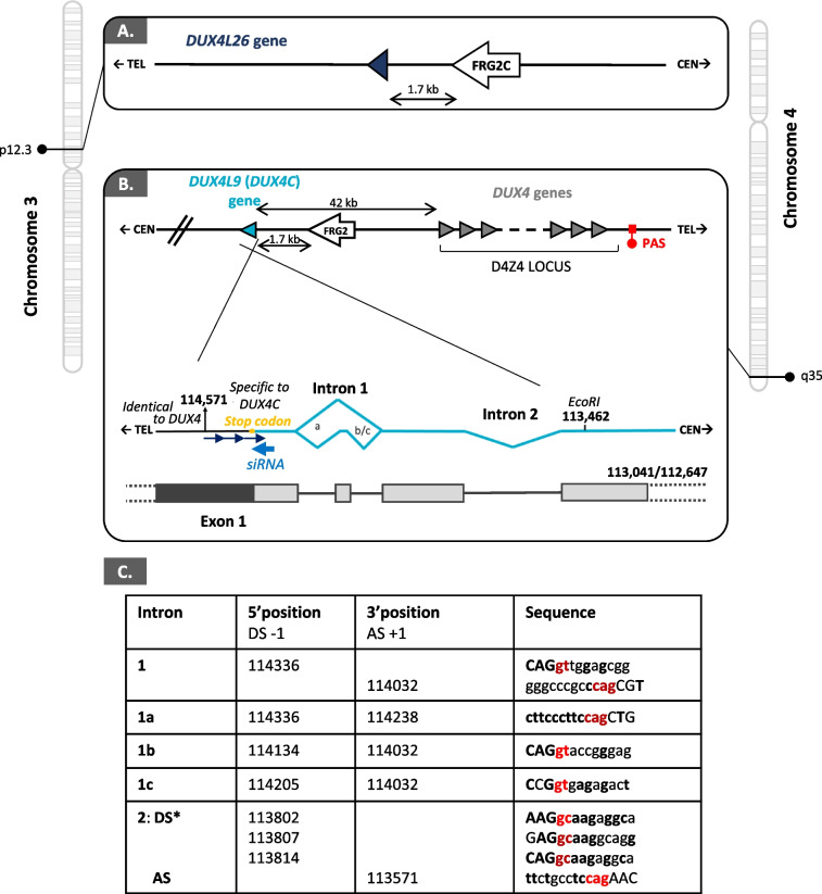
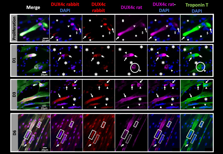
Results: We identified new alternatively spliced DUX4C transcripts and confirmed DUX4c immunodetection in rare FSHD muscle cells in primary culture. DUX4c was detected in nuclei, cytoplasm or at cell-cell contacts between myocytes and interacted sporadically with specific RNA-binding proteins involved, a.o., in muscle differentiation, repair, and mass maintenance. In FSHD muscle sections, DUX4c was found in fibers with unusual shape or central/delocalized nuclei (a regeneration feature) staining for developmental myosin heavy chain, MYOD or presenting intense desmin labeling. Some couples of myocytes/fibers locally exhibited peripheral DUX4c-positive areas that were very close to each other, but in distinct cells. MYOD or intense desmin staining at these locations suggested an imminent muscle cell fusion. We further demonstrated DUX4c interaction with its major protein partner, C1qBP, inside myocytes/myofibers that presented features of regeneration. On adjacent muscle sections, we could unexpectedly detect DUX4 (the FSHD causal protein) and its interaction with C1qBP in fusing myocytes/fibers.
Conclusions: DUX4c upregulation in FSHD muscles suggests it contributes not only to the pathology but also, based on its protein partners and specific markers, to attempts at muscle regeneration. The presence of both DUX4 and DUX4c in regenerating FSHD muscle cells suggests DUX4 could compete with normal DUX4c functions, thus explaining why skeletal muscle is particularly sensitive to DUX4 toxicity. Caution should be exerted with therapeutic agents aiming for DUX4 suppression because they might also repress the highly similar DUX4c and interfere with its physiological role.
期刊介绍:
The only open access journal in its field, Skeletal Muscle publishes novel, cutting-edge research and technological advancements that investigate the molecular mechanisms underlying the biology of skeletal muscle. Reflecting the breadth of research in this area, the journal welcomes manuscripts about the development, metabolism, the regulation of mass and function, aging, degeneration, dystrophy and regeneration of skeletal muscle, with an emphasis on understanding adult skeletal muscle, its maintenance, and its interactions with non-muscle cell types and regulatory modulators.
Main areas of interest include:
-differentiation of skeletal muscle-
atrophy and hypertrophy of skeletal muscle-
aging of skeletal muscle-
regeneration and degeneration of skeletal muscle-
biology of satellite and satellite-like cells-
dystrophic degeneration of skeletal muscle-
energy and glucose homeostasis in skeletal muscle-
non-dystrophic genetic diseases of skeletal muscle, such as Spinal Muscular Atrophy and myopathies-
maintenance of neuromuscular junctions-
roles of ryanodine receptors and calcium signaling in skeletal muscle-
roles of nuclear receptors in skeletal muscle-
roles of GPCRs and GPCR signaling in skeletal muscle-
other relevant aspects of skeletal muscle biology.
In addition, articles on translational clinical studies that address molecular and cellular mechanisms of skeletal muscle will be published. Case reports are also encouraged for submission.
Skeletal Muscle reflects the breadth of research on skeletal muscle and bridges gaps between diverse areas of science for example cardiac cell biology and neurobiology, which share common features with respect to cell differentiation, excitatory membranes, cell-cell communication, and maintenance. Suitable articles are model and mechanism-driven, and apply statistical principles where appropriate; purely descriptive studies are of lesser interest.
 求助内容:
求助内容: 应助结果提醒方式:


