Wenxia Su, Dimeng Zhang, Qiang Zhang, Qianzhong Li
{"title":"Study on the spatial distribution patterns of histone modifications in Hippo pathway genes.","authors":"Wenxia Su, Dimeng Zhang, Qiang Zhang, Qianzhong Li","doi":"10.52601/bpr.2021.200042","DOIUrl":null,"url":null,"abstract":"<p><p>Hippo pathway can regulate cell division, differentiation and apoptosis, and control the shape and size of organs. To study the distribution patterns of histone modifications of Hippo pathway genes in embryonic stem cells is helpful to understand the molecular regulation mechanism of histone modification and Hippo pathway on stem cell self-renewal. In this study, 19 genes of Hippo pathway including YAP, TAZ, LATS1/2, MST1 and SAV1, and eight histone modifications in embryonic stem cells were chosen to study the spatial distribution patterns of histone modifications. It was found that there were obvious type specificity and the location preference of target regions in the distributions of histone modifications, and H3K4me3 and H3K36me3 played the most important regulatory roles. Through the correlation analysis of histone modifications, a histone modification functional cluster composed of H3K4ac, H3K4me3, H3K9ac and H3K27ac was detected in YAP. In addition, the spatial distribution patterns of histone modifications in Hippo pathway genes were obtained, which provided a new theoretical reference for elucidating the mechanism of histone modifications regulating the gene expression of Hippo pathway, and for revealing the molecular regulatory mechanism of histone modifications affecting the self-renewal of embryonic stem cells by regulating the Hippo pathway.</p>","PeriodicalId":59621,"journal":{"name":"生物物理学报:英文版","volume":"7 1","pages":"71-79"},"PeriodicalIF":0.0000,"publicationDate":"2021-02-28","publicationTypes":"Journal Article","fieldsOfStudy":null,"isOpenAccess":false,"openAccessPdf":"https://www.ncbi.nlm.nih.gov/pmc/articles/PMC10240540/pdf/","citationCount":"2","resultStr":null,"platform":"Semanticscholar","paperid":null,"PeriodicalName":"生物物理学报:英文版","FirstCategoryId":"1085","ListUrlMain":"https://doi.org/10.52601/bpr.2021.200042","RegionNum":0,"RegionCategory":null,"ArticlePicture":[],"TitleCN":null,"AbstractTextCN":null,"PMCID":null,"EPubDate":"","PubModel":"","JCR":"","JCRName":"","Score":null,"Total":0}
引用次数: 2
Abstract
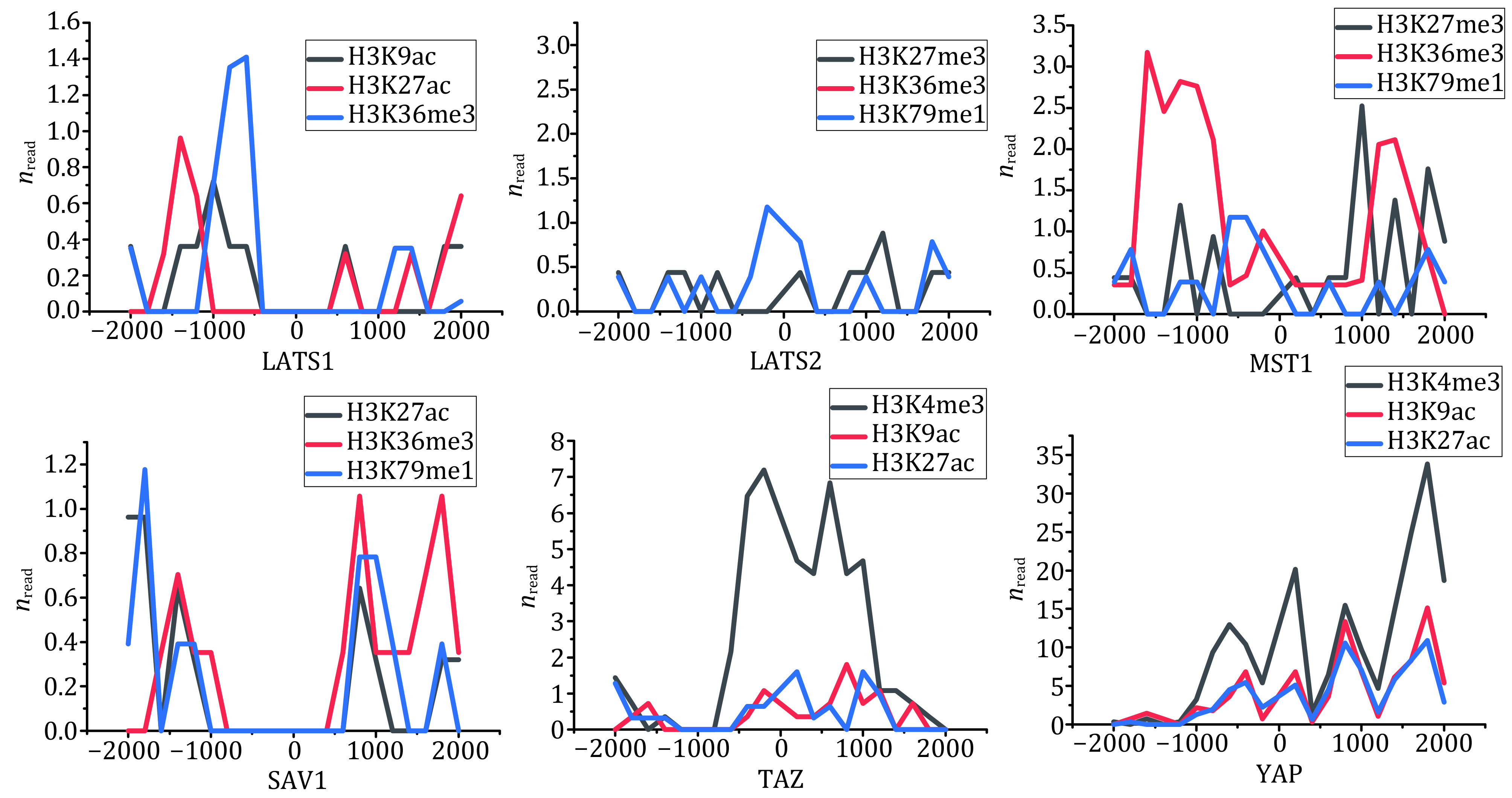
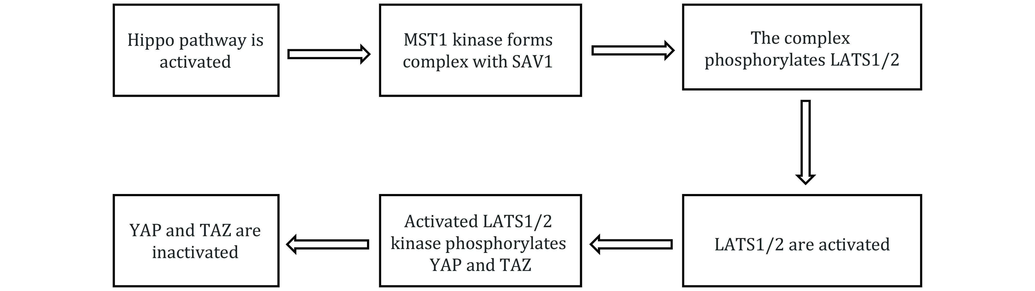
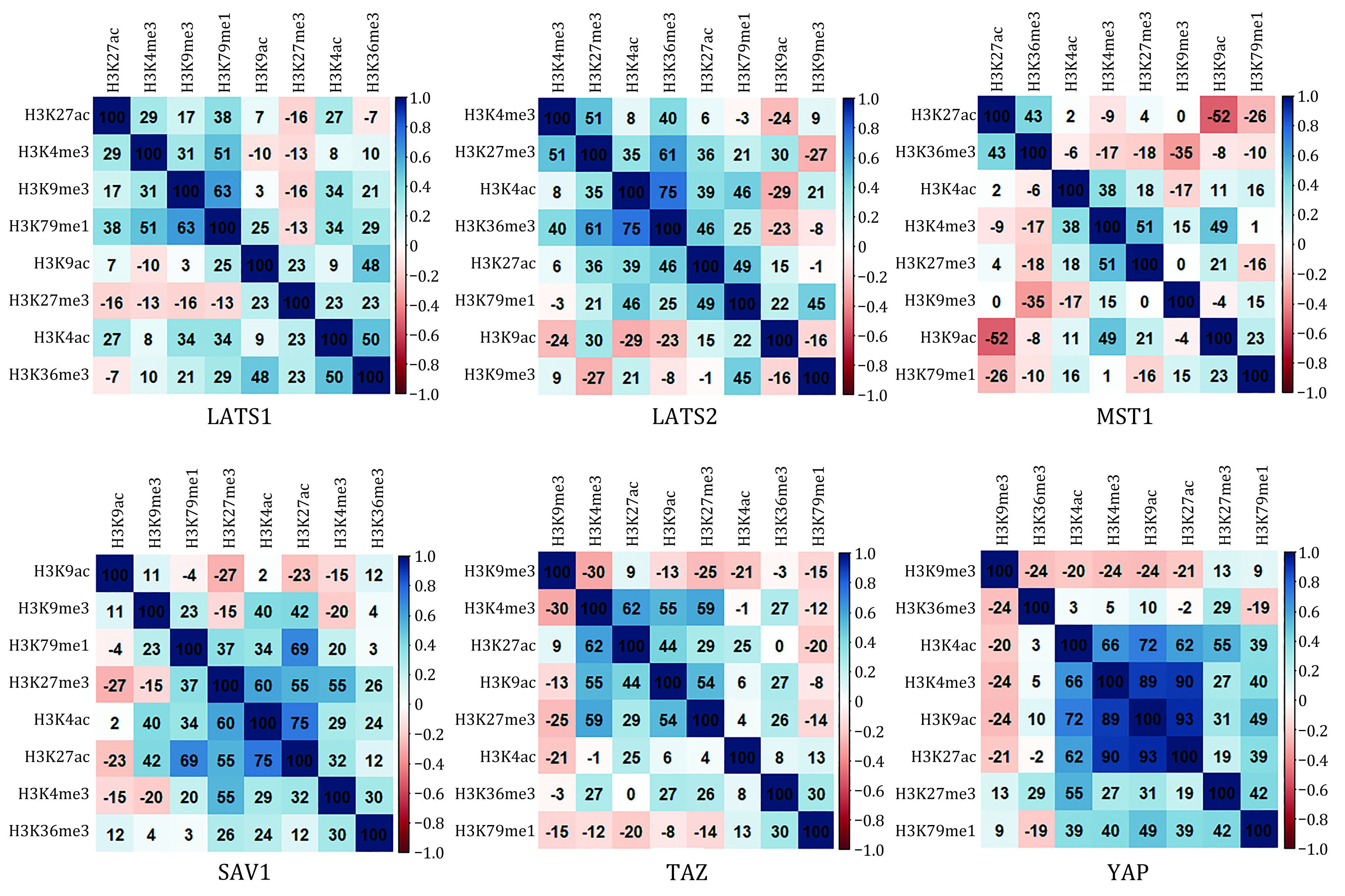
Hippo pathway can regulate cell division, differentiation and apoptosis, and control the shape and size of organs. To study the distribution patterns of histone modifications of Hippo pathway genes in embryonic stem cells is helpful to understand the molecular regulation mechanism of histone modification and Hippo pathway on stem cell self-renewal. In this study, 19 genes of Hippo pathway including YAP, TAZ, LATS1/2, MST1 and SAV1, and eight histone modifications in embryonic stem cells were chosen to study the spatial distribution patterns of histone modifications. It was found that there were obvious type specificity and the location preference of target regions in the distributions of histone modifications, and H3K4me3 and H3K36me3 played the most important regulatory roles. Through the correlation analysis of histone modifications, a histone modification functional cluster composed of H3K4ac, H3K4me3, H3K9ac and H3K27ac was detected in YAP. In addition, the spatial distribution patterns of histone modifications in Hippo pathway genes were obtained, which provided a new theoretical reference for elucidating the mechanism of histone modifications regulating the gene expression of Hippo pathway, and for revealing the molecular regulatory mechanism of histone modifications affecting the self-renewal of embryonic stem cells by regulating the Hippo pathway.
 求助内容:
求助内容: 应助结果提醒方式:


