Dongwoo Hahn, Joseph D Quick, Brian R Thompson, Adelyn Crabtree, Benjamin J Hackel, Frank S Bates, Joseph M Metzger
{"title":"Rapid restitution of contractile dysfunction by synthetic copolymers in dystrophin-deficient single live skeletal muscle fibers.","authors":"Dongwoo Hahn, Joseph D Quick, Brian R Thompson, Adelyn Crabtree, Benjamin J Hackel, Frank S Bates, Joseph M Metzger","doi":"10.1186/s13395-023-00318-y","DOIUrl":null,"url":null,"abstract":"<p><p>Duchenne muscular dystrophy (DMD) is caused by the lack of dystrophin, a cytoskeletal protein essential for the preservation of the structural integrity of the muscle cell membrane. DMD patients develop severe skeletal muscle weakness, degeneration, and early death. We tested here amphiphilic synthetic membrane stabilizers in mdx skeletal muscle fibers (flexor digitorum brevis; FDB) to determine their effectiveness in restoring contractile function in dystrophin-deficient live skeletal muscle fibers. After isolating FDB fibers via enzymatic digestion and trituration from thirty-three adult male mice (9 C57BL10, 24 mdx), these were plated on a laminin-coated coverslip and treated with poloxamer 188 (P188; PEO<sub>75</sub>-PPO<sub>30</sub>-PEO<sub>75</sub>; 8400 g/mol), architecturally inverted triblock (PPO<sub>15</sub>-PEO<sub>200</sub>-PPO<sub>15</sub>, 10,700 g/mol), and diblock (PEO<sub>75</sub>-PPO<sub>16</sub>-C<sub>4</sub>, 4200 g/mol) copolymers. We assessed the twitch kinetics of sarcomere length (SL) and intracellular Ca<sup>2+</sup> transient by Fura-2AM by field stimulation (25 V, 0.2 Hz, 25 °C). Twitch contraction peak SL shortening of mdx FDB fibers was markedly depressed to 30% of the dystrophin-replete control FDB fibers from C57BL10 (P < 0.001). Compared to vehicle-treated mdx FDB fibers, copolymer treatment robustly and rapidly restored the twitch peak SL shortening (all P < 0.05) by P188 (15 μM = + 110%, 150 μM = + 220%), diblock (15 μM = + 50%, 150 μM = + 50%), and inverted triblock copolymer (15 μM = + 180%, 150 μM = + 90%). Twitch peak Ca<sup>2+</sup> transient from mdx FDB fibers was also depressed compared to C57BL10 FDB fibers (P < 0.001). P188 and inverted triblock copolymer treatment of mdx FDB fibers increased the twitch peak Ca<sup>2+</sup> transient (P < 0.001). This study shows synthetic block copolymers with varied architectures can rapidly and highly effectively enhance contractile function in live dystrophin-deficient skeletal muscle fibers.</p>","PeriodicalId":21747,"journal":{"name":"Skeletal Muscle","volume":"13 1","pages":"9"},"PeriodicalIF":4.4000,"publicationDate":"2023-05-19","publicationTypes":"Journal Article","fieldsOfStudy":null,"isOpenAccess":false,"openAccessPdf":"https://www.ncbi.nlm.nih.gov/pmc/articles/PMC10197332/pdf/","citationCount":"0","resultStr":null,"platform":"Semanticscholar","paperid":null,"PeriodicalName":"Skeletal Muscle","FirstCategoryId":"3","ListUrlMain":"https://doi.org/10.1186/s13395-023-00318-y","RegionNum":2,"RegionCategory":"医学","ArticlePicture":[],"TitleCN":null,"AbstractTextCN":null,"PMCID":null,"EPubDate":"","PubModel":"","JCR":"Q2","JCRName":"CELL BIOLOGY","Score":null,"Total":0}
引用次数: 0
Abstract
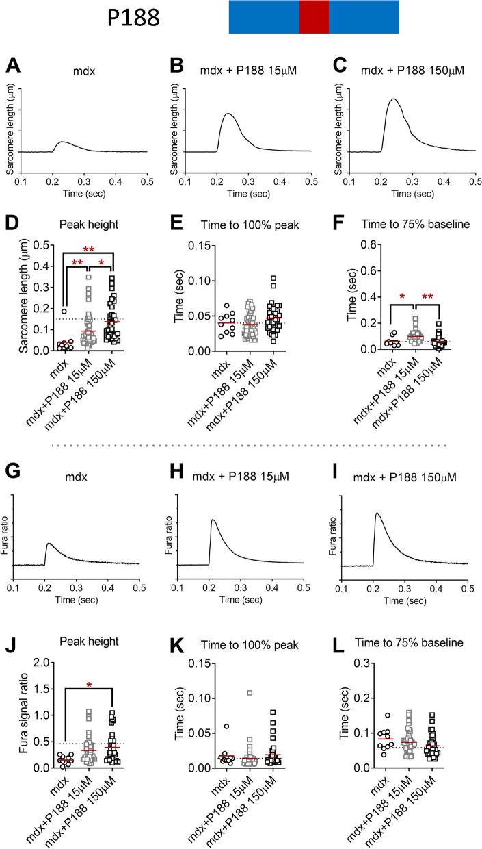
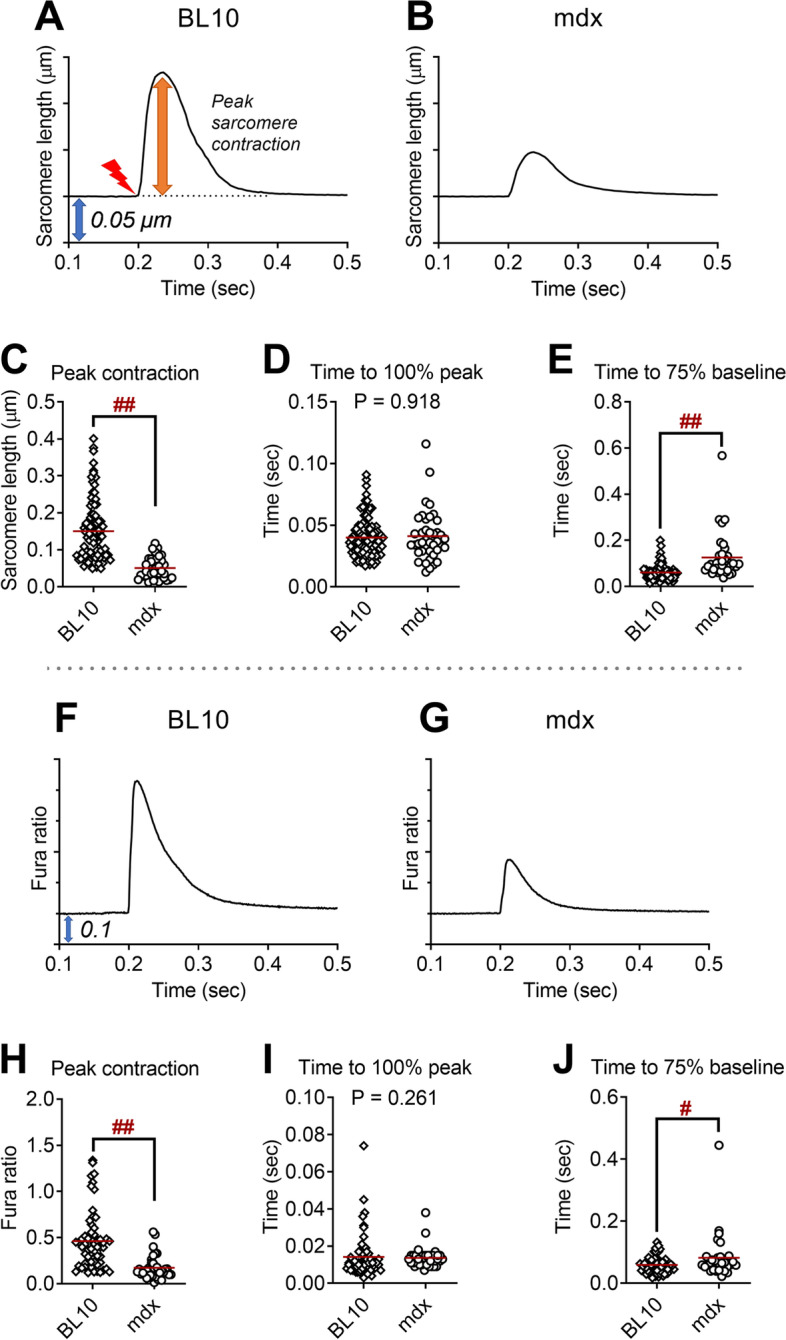
Duchenne muscular dystrophy (DMD) is caused by the lack of dystrophin, a cytoskeletal protein essential for the preservation of the structural integrity of the muscle cell membrane. DMD patients develop severe skeletal muscle weakness, degeneration, and early death. We tested here amphiphilic synthetic membrane stabilizers in mdx skeletal muscle fibers (flexor digitorum brevis; FDB) to determine their effectiveness in restoring contractile function in dystrophin-deficient live skeletal muscle fibers. After isolating FDB fibers via enzymatic digestion and trituration from thirty-three adult male mice (9 C57BL10, 24 mdx), these were plated on a laminin-coated coverslip and treated with poloxamer 188 (P188; PEO75-PPO30-PEO75; 8400 g/mol), architecturally inverted triblock (PPO15-PEO200-PPO15, 10,700 g/mol), and diblock (PEO75-PPO16-C4, 4200 g/mol) copolymers. We assessed the twitch kinetics of sarcomere length (SL) and intracellular Ca2+ transient by Fura-2AM by field stimulation (25 V, 0.2 Hz, 25 °C). Twitch contraction peak SL shortening of mdx FDB fibers was markedly depressed to 30% of the dystrophin-replete control FDB fibers from C57BL10 (P < 0.001). Compared to vehicle-treated mdx FDB fibers, copolymer treatment robustly and rapidly restored the twitch peak SL shortening (all P < 0.05) by P188 (15 μM = + 110%, 150 μM = + 220%), diblock (15 μM = + 50%, 150 μM = + 50%), and inverted triblock copolymer (15 μM = + 180%, 150 μM = + 90%). Twitch peak Ca2+ transient from mdx FDB fibers was also depressed compared to C57BL10 FDB fibers (P < 0.001). P188 and inverted triblock copolymer treatment of mdx FDB fibers increased the twitch peak Ca2+ transient (P < 0.001). This study shows synthetic block copolymers with varied architectures can rapidly and highly effectively enhance contractile function in live dystrophin-deficient skeletal muscle fibers.
期刊介绍:
The only open access journal in its field, Skeletal Muscle publishes novel, cutting-edge research and technological advancements that investigate the molecular mechanisms underlying the biology of skeletal muscle. Reflecting the breadth of research in this area, the journal welcomes manuscripts about the development, metabolism, the regulation of mass and function, aging, degeneration, dystrophy and regeneration of skeletal muscle, with an emphasis on understanding adult skeletal muscle, its maintenance, and its interactions with non-muscle cell types and regulatory modulators.
Main areas of interest include:
-differentiation of skeletal muscle-
atrophy and hypertrophy of skeletal muscle-
aging of skeletal muscle-
regeneration and degeneration of skeletal muscle-
biology of satellite and satellite-like cells-
dystrophic degeneration of skeletal muscle-
energy and glucose homeostasis in skeletal muscle-
non-dystrophic genetic diseases of skeletal muscle, such as Spinal Muscular Atrophy and myopathies-
maintenance of neuromuscular junctions-
roles of ryanodine receptors and calcium signaling in skeletal muscle-
roles of nuclear receptors in skeletal muscle-
roles of GPCRs and GPCR signaling in skeletal muscle-
other relevant aspects of skeletal muscle biology.
In addition, articles on translational clinical studies that address molecular and cellular mechanisms of skeletal muscle will be published. Case reports are also encouraged for submission.
Skeletal Muscle reflects the breadth of research on skeletal muscle and bridges gaps between diverse areas of science for example cardiac cell biology and neurobiology, which share common features with respect to cell differentiation, excitatory membranes, cell-cell communication, and maintenance. Suitable articles are model and mechanism-driven, and apply statistical principles where appropriate; purely descriptive studies are of lesser interest.
 求助内容:
求助内容: 应助结果提醒方式:


