Chaos analysis of the cortical boundary for the recognition of psychosis.

IF 4.1 2区 医学 Q2 NEUROSCIENCES
Alexandra I Korda, Christina Andreou, Mihai Avram, Marina Frisman, Mariya Aliqadri, Anita Riecher-Rössler, Heinz Handels, Thomas Martinetz, Stefan Borgwardt
{"title":"Chaos analysis of the cortical boundary for the recognition of psychosis.","authors":"Alexandra I Korda, Christina Andreou, Mihai Avram, Marina Frisman, Mariya Aliqadri, Anita Riecher-Rössler, Heinz Handels, Thomas Martinetz, Stefan Borgwardt","doi":"10.1503/jpn.220160","DOIUrl":null,"url":null,"abstract":"Background: Structural MRI studies in people with first-episode psychosis (FEP) and those in the clinical high-risk (CHR) state have consistently shown volumetric abnormalities that depict changes in the structural complexity of the cortical boundary. The aim of the present study was to employ chaos analysis in the identification of people with psychosis based on the structural complexity of the cortical boundary and subcortical areas. Methods: We performed chaos analysis of the grey matter distribution on structural MRIs. First, the outer boundary points for each slice in the axial, coronal and sagittal view were calculated for grey matter maps. Next, the distance of each boundary point from the centre of mass in the grey matter was calculated and stored as spatial series, which was further analyzed by extracting the Largest Lyapunov Exponent (lambda [λ]), a feature depicting the structural complexity of the cortical boundary. Results: Structural MRIs were acquired from 77 FEP, 73 CHR and 44 healthy controls. We compared λ brain maps between groups, which resulted in statistically significant differences in all comparisons. By matching the λ values extracted in axial view with the Morlet wavelet, differences on the surface relief are observed between groups. Limitations: Parameters were selected after experimentation on the examined sample. Investigation of the effectiveness of the method in a larger data set is needed. Conclusion: The proposed framework using spatial series verifies diagnosis-relevant features and may contribute to the identification of structural biomarkers for psychosis.","PeriodicalId":50073,"journal":{"name":"Journal of Psychiatry & Neuroscience","volume":"48 2","pages":"E135-E142"},"PeriodicalIF":4.1000,"publicationDate":"2023-03-01","publicationTypes":"Journal Article","fieldsOfStudy":null,"isOpenAccess":false,"openAccessPdf":"https://ftp.ncbi.nlm.nih.gov/pub/pmc/oa_pdf/7f/02/48-2-E135.PMC10139064.pdf","citationCount":"0","resultStr":null,"platform":"Semanticscholar","paperid":null,"PeriodicalName":"Journal of Psychiatry & Neuroscience","FirstCategoryId":"3","ListUrlMain":"https://doi.org/10.1503/jpn.220160","RegionNum":2,"RegionCategory":"医学","ArticlePicture":[],"TitleCN":null,"AbstractTextCN":null,"PMCID":null,"EPubDate":"","PubModel":"","JCR":"Q2","JCRName":"NEUROSCIENCES","Score":null,"Total":0}
引用次数: 0

Abstract

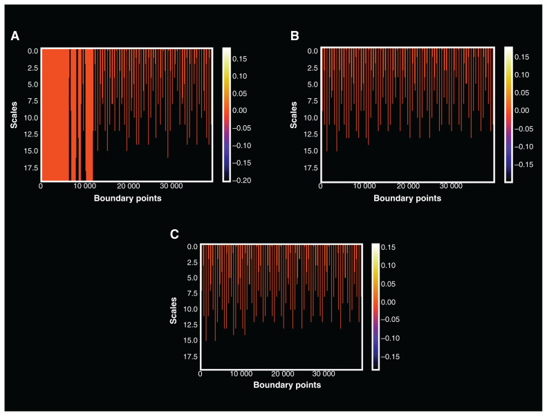
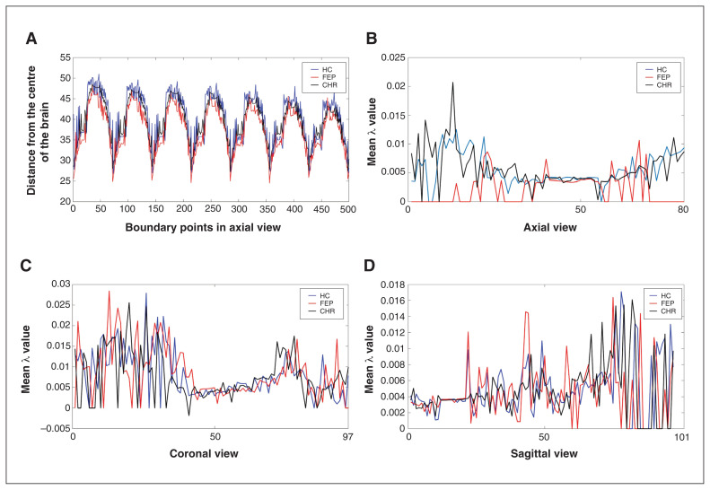
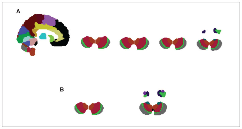
Background: Structural MRI studies in people with first-episode psychosis (FEP) and those in the clinical high-risk (CHR) state have consistently shown volumetric abnormalities that depict changes in the structural complexity of the cortical boundary. The aim of the present study was to employ chaos analysis in the identification of people with psychosis based on the structural complexity of the cortical boundary and subcortical areas. Methods: We performed chaos analysis of the grey matter distribution on structural MRIs. First, the outer boundary points for each slice in the axial, coronal and sagittal view were calculated for grey matter maps. Next, the distance of each boundary point from the centre of mass in the grey matter was calculated and stored as spatial series, which was further analyzed by extracting the Largest Lyapunov Exponent (lambda [λ]), a feature depicting the structural complexity of the cortical boundary. Results: Structural MRIs were acquired from 77 FEP, 73 CHR and 44 healthy controls. We compared λ brain maps between groups, which resulted in statistically significant differences in all comparisons. By matching the λ values extracted in axial view with the Morlet wavelet, differences on the surface relief are observed between groups. Limitations: Parameters were selected after experimentation on the examined sample. Investigation of the effectiveness of the method in a larger data set is needed. Conclusion: The proposed framework using spatial series verifies diagnosis-relevant features and may contribute to the identification of structural biomarkers for psychosis.
Abstract Image

Abstract Image

Abstract Image

大脑皮层边界混沌分析对精神病的识别。
背景:对首发精神病(FEP)患者和临床高危(CHR)患者的结构MRI研究一致显示体积异常,描绘了皮质边界结构复杂性的变化。本研究的目的是基于皮层边界和皮层下区域的结构复杂性,运用混沌分析来识别精神病患者。方法:对结构核磁共振成像的灰质分布进行混沌分析。首先,对灰质图进行轴向、冠状和矢状视图各切片的外边界点计算;接下来,计算每个边界点到灰质质心的距离并将其存储为空间序列,通过提取最大李雅普诺夫指数(λ [λ])进一步分析,该指数是描述皮质边界结构复杂性的特征。结果:FEP 77例,CHR 73例,健康对照44例。我们比较了各组之间的λ脑图,结果在所有比较中都有统计学上的显著差异。将轴向视场提取的λ值与Morlet小波进行匹配,观察各组地表起伏的差异。局限性:参数是在检测样品上实验后选择的。需要在更大的数据集上研究该方法的有效性。结论:使用空间序列的框架验证了诊断相关特征,并可能有助于识别精神病的结构生物标志物。
本文章由计算机程序翻译,如有差异,请以英文原文为准。
求助全文
约1分钟内获得全文 求助全文
来源期刊
CiteScore
6.80
自引率
2.30%
发文量
51
审稿时长
2 months
期刊介绍: The Journal of Psychiatry & Neuroscience publishes papers at the intersection of psychiatry and neuroscience that advance our understanding of the neural mechanisms involved in the etiology and treatment of psychiatric disorders. This includes studies on patients with psychiatric disorders, healthy humans, and experimental animals as well as studies in vitro. Original research articles, including clinical trials with a mechanistic component, and review papers will be considered.
×
引用
GB/T 7714-2015
复制
MLA
复制
APA
复制
导出至
BibTeX EndNote RefMan NoteFirst NoteExpress
×
提示
您的信息不完整,为了账户安全,请先补充。
现在去补充
×
提示
您因"违规操作"
具体请查看互助需知
我知道了
×
提示
确定
请完成安全验证×
copy
已复制链接
快去分享给好友吧!
我知道了
右上角分享
点击右上角分享
0
联系我们:info@booksci.cn Book学术提供免费学术资源搜索服务,方便国内外学者检索中英文文献。致力于提供最便捷和优质的服务体验。 Copyright © 2023 布克学术 All rights reserved.
京ICP备2023020795号-1
ghs 京公网安备 11010802042870号
Book学术文献互助
Book学术文献互助群
群 号:481959085
Book学术官方微信