Yao Liu , Zhenzhen Xun , Kun Ma , Shuhang Liang , Xianying Li , Shuo Zhou , Linmao Sun , Yufeng Liu , Yanhua Du , Xinyu Guo , Tianming Cui , Huanran Zhou , Jizhou Wang , Dalong Yin , Ruipeng Song , Shugeng Zhang , Wei Cai , Fanzheng Meng , Hongrui Guo , Bo Zhang , Lianxin Liu
{"title":"Identification of a tumour immune barrier in the HCC microenvironment that determines the efficacy of immunotherapy","authors":"Yao Liu , Zhenzhen Xun , Kun Ma , Shuhang Liang , Xianying Li , Shuo Zhou , Linmao Sun , Yufeng Liu , Yanhua Du , Xinyu Guo , Tianming Cui , Huanran Zhou , Jizhou Wang , Dalong Yin , Ruipeng Song , Shugeng Zhang , Wei Cai , Fanzheng Meng , Hongrui Guo , Bo Zhang , Lianxin Liu","doi":"10.1016/j.jhep.2023.01.011","DOIUrl":null,"url":null,"abstract":"<div><h3>Background & Aims</h3><p><span>The tumour microenvironment<span> (TME) is a crucial mediator of cancer progression and therapeutic outcome. The TME subtype correlates with patient response to immunotherapy in </span></span>multiple cancers. Most previous studies have focused on the role of different cellular components in the TME associated with immunotherapy efficacy. However, the specific structure of the TME and its role in immunotherapy efficacy remain largely unknown.</p></div><div><h3>Methods</h3><p><span>We combined spatial transcriptomics<span> with single-cell RNA-sequencing and multiplexed immunofluorescence to identify the specific spatial structures in the TME that determine the efficacy of immunotherapy </span></span>in patients<span> with hepatocellular carcinoma (HCC) receiving anti-PD-1 treatment.</span></p></div><div><h3>Results</h3><p>We identified a tumour immune barrier (TIB) structure, a spatial niche composed of <em>SPP1</em><sup><em>+</em></sup><span> macrophages and cancer-associated fibroblasts (CAFs) located near the tumour boundary, which is associated with the efficacy of immune checkpoint blockade. Furthermore, we dissected ligand‒receptor networks among malignant cells, </span><em>SPP1</em><sup><em>+</em></sup><span> macrophages, and CAFs; that is, the hypoxic microenvironment promotes SPP1 expression, and </span><em>SPP1</em><sup><em>+</em></sup><span> macrophages interact with CAFs to stimulate extracellular matrix remodelling and promote TIB structure formation, thereby limiting immune infiltration in the tumour core. Preclinically, the blockade of SPP1 or macrophage-specific deletion of </span><em>Spp1</em> in mice led to enhanced efficacy of anti-PD-1 treatment in mouse liver cancer, accompanied by reduced CAF infiltration and increased cytotoxic T-cell infiltration.</p></div><div><h3>Conclusions</h3><p>We identified that the TIB structure formed by the interaction of <em>SPP1</em><sup><em>+</em></sup> macrophages and CAFs is related to immunotherapy efficacy. Therefore, disruption of the TIB structure by blocking SPP1 may be considered a relevant therapeutic approach to enhance the therapeutic effect of immune checkpoint blockade in HCC.</p></div><div><h3>Impact and implications</h3><p><span><span>Only a limited number of patients with hepatocellular carcinoma (HCC) benefit from tumour immunotherapy, which significantly hinders its application. Herein, we used multiomics to identify the spatial structure of the tumour immune barrier (TIB), which is formed by the interaction of SPP1+ macrophages and cancer-associated fibroblasts in the HCC microenvironment. This structure constrains immunotherapy efficacy by limiting </span>immune cell infiltration into malignant regions. Preclinically, we revealed that blocking SPP1 or macrophage-specific deletion of </span><em>Spp1</em><span><span> in mice could destroy the TIB structure and sensitize HCC cells to immunotherapy. These results provide the first key steps towards finding more effective therapies for HCC and have implications for physicians, scientists, and </span>drug developers in the field of HCC.</span></p></div>","PeriodicalId":15888,"journal":{"name":"Journal of Hepatology","volume":"78 4","pages":"Pages 770-782"},"PeriodicalIF":26.8000,"publicationDate":"2023-04-01","publicationTypes":"Journal Article","fieldsOfStudy":null,"isOpenAccess":false,"openAccessPdf":"","citationCount":"47","resultStr":null,"platform":"Semanticscholar","paperid":null,"PeriodicalName":"Journal of Hepatology","FirstCategoryId":"3","ListUrlMain":"https://www.sciencedirect.com/science/article/pii/S0168827823000235","RegionNum":1,"RegionCategory":"医学","ArticlePicture":[],"TitleCN":null,"AbstractTextCN":null,"PMCID":null,"EPubDate":"","PubModel":"","JCR":"Q1","JCRName":"GASTROENTEROLOGY & HEPATOLOGY","Score":null,"Total":0}
引用次数: 47
Abstract
Background & Aims
The tumour microenvironment (TME) is a crucial mediator of cancer progression and therapeutic outcome. The TME subtype correlates with patient response to immunotherapy in multiple cancers. Most previous studies have focused on the role of different cellular components in the TME associated with immunotherapy efficacy. However, the specific structure of the TME and its role in immunotherapy efficacy remain largely unknown.
Methods
We combined spatial transcriptomics with single-cell RNA-sequencing and multiplexed immunofluorescence to identify the specific spatial structures in the TME that determine the efficacy of immunotherapy in patients with hepatocellular carcinoma (HCC) receiving anti-PD-1 treatment.
Results
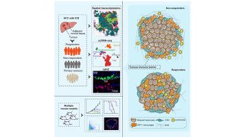
We identified a tumour immune barrier (TIB) structure, a spatial niche composed of SPP1+ macrophages and cancer-associated fibroblasts (CAFs) located near the tumour boundary, which is associated with the efficacy of immune checkpoint blockade. Furthermore, we dissected ligand‒receptor networks among malignant cells, SPP1+ macrophages, and CAFs; that is, the hypoxic microenvironment promotes SPP1 expression, and SPP1+ macrophages interact with CAFs to stimulate extracellular matrix remodelling and promote TIB structure formation, thereby limiting immune infiltration in the tumour core. Preclinically, the blockade of SPP1 or macrophage-specific deletion of Spp1 in mice led to enhanced efficacy of anti-PD-1 treatment in mouse liver cancer, accompanied by reduced CAF infiltration and increased cytotoxic T-cell infiltration.
Conclusions
We identified that the TIB structure formed by the interaction of SPP1+ macrophages and CAFs is related to immunotherapy efficacy. Therefore, disruption of the TIB structure by blocking SPP1 may be considered a relevant therapeutic approach to enhance the therapeutic effect of immune checkpoint blockade in HCC.
Impact and implications
Only a limited number of patients with hepatocellular carcinoma (HCC) benefit from tumour immunotherapy, which significantly hinders its application. Herein, we used multiomics to identify the spatial structure of the tumour immune barrier (TIB), which is formed by the interaction of SPP1+ macrophages and cancer-associated fibroblasts in the HCC microenvironment. This structure constrains immunotherapy efficacy by limiting immune cell infiltration into malignant regions. Preclinically, we revealed that blocking SPP1 or macrophage-specific deletion of Spp1 in mice could destroy the TIB structure and sensitize HCC cells to immunotherapy. These results provide the first key steps towards finding more effective therapies for HCC and have implications for physicians, scientists, and drug developers in the field of HCC.
期刊介绍:
The Journal of Hepatology is the official publication of the European Association for the Study of the Liver (EASL). It is dedicated to presenting clinical and basic research in the field of hepatology through original papers, reviews, case reports, and letters to the Editor. The Journal is published in English and may consider supplements that pass an editorial review.
 求助内容:
求助内容: 应助结果提醒方式:


