Han-Sol Lee, Kyu-Young Oh, Ju-Hee Kang, Jo-Eun Kim, Kyung-Hoe Huh, Won-Jin Yi, Min-Suk Heo, Sam-Sun Lee
{"title":"A case report of an unusual temporomandibular joint mass: Nodular fasciitis.","authors":"Han-Sol Lee, Kyu-Young Oh, Ju-Hee Kang, Jo-Eun Kim, Kyung-Hoe Huh, Won-Jin Yi, Min-Suk Heo, Sam-Sun Lee","doi":"10.5624/isd.20220175","DOIUrl":null,"url":null,"abstract":"<p><p>Nodular fasciitis (NF) is a benign myofibroblastic proliferation that grows very rapidly, mimicking a sarcoma on imaging. It is treated by local excision, and recurrence has been reported in only a few cases, even when excised incompletely. The most prevalent diagnoses of temporomandibular joint (TMJ) masses include synovial chondromatosis, pigmented villonodular synovitis, and sarcomas. Cases of NF in the TMJ are extremely rare, and only 3 cases have been reported to date. Due to its destructive features and rarity, NF has often been misdiagnosed as a more aggressive lesion, which could expose patients to unnecessary and invasive treatment approaches beyond repair. This report presents a case of NF in the TMJ, focusing on various imaging features, along with a literature review aiming to determine the hallmark features of NF in the TMJ and highlight the diagnostic challenges.</p>","PeriodicalId":51714,"journal":{"name":"Imaging Science in Dentistry","volume":"53 1","pages":"83-89"},"PeriodicalIF":1.7000,"publicationDate":"2023-03-01","publicationTypes":"Journal Article","fieldsOfStudy":null,"isOpenAccess":false,"openAccessPdf":"https://ftp.ncbi.nlm.nih.gov/pub/pmc/oa_pdf/e4/23/isd-53-83.PMC10060757.pdf","citationCount":"0","resultStr":null,"platform":"Semanticscholar","paperid":null,"PeriodicalName":"Imaging Science in Dentistry","FirstCategoryId":"1085","ListUrlMain":"https://doi.org/10.5624/isd.20220175","RegionNum":0,"RegionCategory":null,"ArticlePicture":[],"TitleCN":null,"AbstractTextCN":null,"PMCID":null,"EPubDate":"","PubModel":"","JCR":"Q3","JCRName":"DENTISTRY, ORAL SURGERY & MEDICINE","Score":null,"Total":0}
引用次数: 0
Abstract
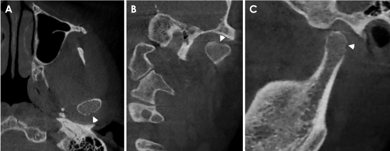
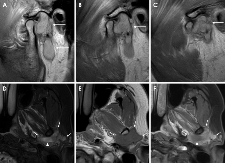
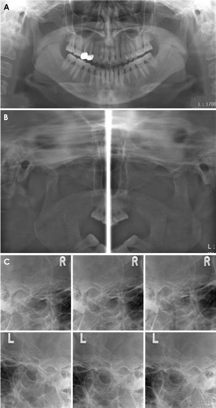
Nodular fasciitis (NF) is a benign myofibroblastic proliferation that grows very rapidly, mimicking a sarcoma on imaging. It is treated by local excision, and recurrence has been reported in only a few cases, even when excised incompletely. The most prevalent diagnoses of temporomandibular joint (TMJ) masses include synovial chondromatosis, pigmented villonodular synovitis, and sarcomas. Cases of NF in the TMJ are extremely rare, and only 3 cases have been reported to date. Due to its destructive features and rarity, NF has often been misdiagnosed as a more aggressive lesion, which could expose patients to unnecessary and invasive treatment approaches beyond repair. This report presents a case of NF in the TMJ, focusing on various imaging features, along with a literature review aiming to determine the hallmark features of NF in the TMJ and highlight the diagnostic challenges.

 求助内容:
求助内容: 应助结果提醒方式:


