Han Song, Gang Zheng, Xue-Feng Shen, Zai-Hua Zhao, Yang Liu, Yang Liu, Ying-Ying Liu, Jun-Jun Kang, Jing-Yuan Chen, Wen-Jing Luo
{"title":"An efficient autometallography approach to localize lead at ultra-structural levels of cultured cells.","authors":"Han Song, Gang Zheng, Xue-Feng Shen, Zai-Hua Zhao, Yang Liu, Yang Liu, Ying-Ying Liu, Jun-Jun Kang, Jing-Yuan Chen, Wen-Jing Luo","doi":"10.1007/s41048-020-00116-9","DOIUrl":null,"url":null,"abstract":"<p><p>Understanding the precise intracellular localization of lead (Pb) is a key in deciphering processes in Pb-induced toxicology. However, it is a great challenge to trace Pb <i>in vitro</i>, especially in cultured cells. We aimed to find an innovative and efficient approach to investigate distribution of Pb in cells and to validate it through determining the subcellular Pb content. We identified its ultra-structural distribution with autometallography under electron microscopy in a choroidal epithelial Z310 cell line. Electron microscopy in combination with energy-dispersive X-ray spectroscope (EDS) was employed to provide further evidence of Pb location. In addition, Pb content was determined in the cytosol, membrane/organelle, nucleus and cytoskeleton fractions with atomic absorption spectroscopy. Pb was found predominantly inside the nuclear membranes and some was distributed in the cytoplasm under low-concentration exposure. Nuclear existence of Pb was verified by EDS under electron microscopy. Once standardized for protein content, Pb percentage in the nucleus fraction reached the highest level (76%). Our results indicate that Pb is accumulated mainly in the nucleus of choroid plexus. This method is sensitive and precise in providing optimal means to study the ultra-structural localization of Pb for <i>in vitro</i> models. In addition, it offers the possibility of localization of other metals in cultured cells. Some procedures may also be adopted to detect target proteins via immunoreactions.</p>","PeriodicalId":59621,"journal":{"name":"生物物理学报:英文版","volume":"6 5","pages":"211-221"},"PeriodicalIF":0.0000,"publicationDate":"2020-10-31","publicationTypes":"Journal Article","fieldsOfStudy":null,"isOpenAccess":false,"openAccessPdf":"https://sci-hub-pdf.com/10.1007/s41048-020-00116-9","citationCount":"0","resultStr":null,"platform":"Semanticscholar","paperid":null,"PeriodicalName":"生物物理学报:英文版","FirstCategoryId":"1085","ListUrlMain":"https://doi.org/10.1007/s41048-020-00116-9","RegionNum":0,"RegionCategory":null,"ArticlePicture":[],"TitleCN":null,"AbstractTextCN":null,"PMCID":null,"EPubDate":"","PubModel":"","JCR":"","JCRName":"","Score":null,"Total":0}
引用次数: 0
Abstract
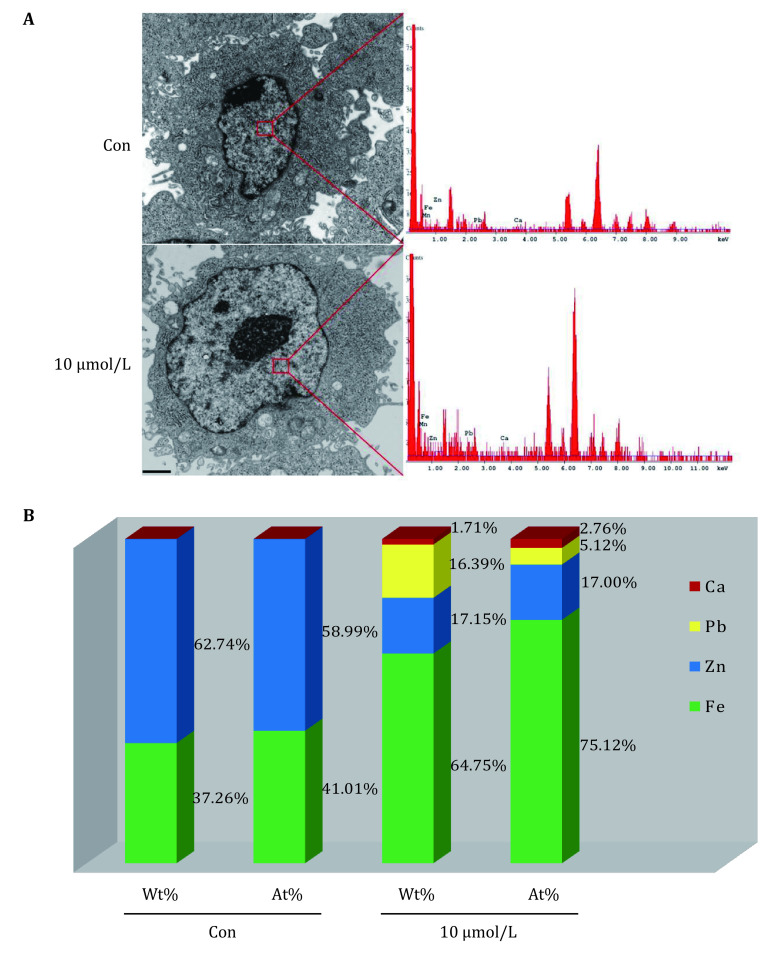
Understanding the precise intracellular localization of lead (Pb) is a key in deciphering processes in Pb-induced toxicology. However, it is a great challenge to trace Pb in vitro, especially in cultured cells. We aimed to find an innovative and efficient approach to investigate distribution of Pb in cells and to validate it through determining the subcellular Pb content. We identified its ultra-structural distribution with autometallography under electron microscopy in a choroidal epithelial Z310 cell line. Electron microscopy in combination with energy-dispersive X-ray spectroscope (EDS) was employed to provide further evidence of Pb location. In addition, Pb content was determined in the cytosol, membrane/organelle, nucleus and cytoskeleton fractions with atomic absorption spectroscopy. Pb was found predominantly inside the nuclear membranes and some was distributed in the cytoplasm under low-concentration exposure. Nuclear existence of Pb was verified by EDS under electron microscopy. Once standardized for protein content, Pb percentage in the nucleus fraction reached the highest level (76%). Our results indicate that Pb is accumulated mainly in the nucleus of choroid plexus. This method is sensitive and precise in providing optimal means to study the ultra-structural localization of Pb for in vitro models. In addition, it offers the possibility of localization of other metals in cultured cells. Some procedures may also be adopted to detect target proteins via immunoreactions.
 求助内容:
求助内容: 应助结果提醒方式:


