Lino Piotto, Roger Gent, Ajay Taranath, Giovanni Bibbo, Day Way Goh
{"title":"Ultrasound diagnosis of hypertrophic pyloric stenosis – Time to change the criteria","authors":"Lino Piotto, Roger Gent, Ajay Taranath, Giovanni Bibbo, Day Way Goh","doi":"10.1002/ajum.12305","DOIUrl":null,"url":null,"abstract":"<div>\n \n \n <section>\n \n <h3> Introduction</h3>\n \n <p>Ultrasound is the examination of choice for the diagnosis of hypertrophic pyloric stenosis (HPS). A correct diagnosis is dependent on the technique and measurement accuracy. However, in the world literature there is a wide range of values suggested for the diagnosis of this condition. The current minimum measurements used to diagnose HPS seem excessively large, and therefore, we set out to redefine these values.</p>\n </section>\n \n <section>\n \n <h3> Methods</h3>\n \n <p>A retrospective study was performed on 607 patients (615 scans) being investigated for HPS. The length and transverse diameter of the pyloric canal, and thickness of the pyloric muscle were measured. All results were correlated with clinical and surgical findings.</p>\n </section>\n \n <section>\n \n <h3> Results</h3>\n \n <p>In this study, the muscle thickness in the normal group was <2.0 mm than in HPS infants having a muscle thickness of 2.0–5.0 mm. All the pyloric canal lengths in the normal group were <5.0 mm than in those with HPS having a length of 10.0–24.0 mm. The transverse diameters ranged from 6.0 to 11.0 mm in the normal group compared with those with HPS having a diameter between 8.0 and 16.0 mm.</p>\n </section>\n \n <section>\n \n <h3> Conclusions</h3>\n \n <p>The current criteria for sonographic diagnosis of HPS should be redefined. The canal length is the single most important discriminator, with a clear separation between normal and abnormal. The commonly used 16.0-mm measurement is too long and should be reduced to 10.0 mm (without the risk of false positives). In many cases, the muscle thickness in those with HPS is as low as 2.0 mm, considerably less than the 3.0 mm that is currently used. The transverse diameter is not a useful discriminator for HPS. The use of current values will delay the diagnosis and timely treatment of this condition.</p>\n </section>\n </div>","PeriodicalId":36517,"journal":{"name":"Australasian Journal of Ultrasound in Medicine","volume":"25 3","pages":"116-126"},"PeriodicalIF":0.0000,"publicationDate":"2022-06-10","publicationTypes":"Journal Article","fieldsOfStudy":null,"isOpenAccess":false,"openAccessPdf":"https://www.ncbi.nlm.nih.gov/pmc/articles/PMC9351432/pdf/AJUM-25-116.pdf","citationCount":"3","resultStr":null,"platform":"Semanticscholar","paperid":null,"PeriodicalName":"Australasian Journal of Ultrasound in Medicine","FirstCategoryId":"1085","ListUrlMain":"https://onlinelibrary.wiley.com/doi/10.1002/ajum.12305","RegionNum":0,"RegionCategory":null,"ArticlePicture":[],"TitleCN":null,"AbstractTextCN":null,"PMCID":null,"EPubDate":"","PubModel":"","JCR":"Q3","JCRName":"Medicine","Score":null,"Total":0}
引用次数: 3
Abstract
Introduction
Ultrasound is the examination of choice for the diagnosis of hypertrophic pyloric stenosis (HPS). A correct diagnosis is dependent on the technique and measurement accuracy. However, in the world literature there is a wide range of values suggested for the diagnosis of this condition. The current minimum measurements used to diagnose HPS seem excessively large, and therefore, we set out to redefine these values.
Methods
A retrospective study was performed on 607 patients (615 scans) being investigated for HPS. The length and transverse diameter of the pyloric canal, and thickness of the pyloric muscle were measured. All results were correlated with clinical and surgical findings.
Results
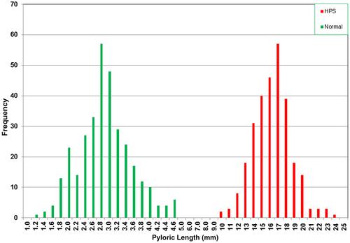
In this study, the muscle thickness in the normal group was <2.0 mm than in HPS infants having a muscle thickness of 2.0–5.0 mm. All the pyloric canal lengths in the normal group were <5.0 mm than in those with HPS having a length of 10.0–24.0 mm. The transverse diameters ranged from 6.0 to 11.0 mm in the normal group compared with those with HPS having a diameter between 8.0 and 16.0 mm.
Conclusions
The current criteria for sonographic diagnosis of HPS should be redefined. The canal length is the single most important discriminator, with a clear separation between normal and abnormal. The commonly used 16.0-mm measurement is too long and should be reduced to 10.0 mm (without the risk of false positives). In many cases, the muscle thickness in those with HPS is as low as 2.0 mm, considerably less than the 3.0 mm that is currently used. The transverse diameter is not a useful discriminator for HPS. The use of current values will delay the diagnosis and timely treatment of this condition.