{"title":"Cone-beam computed tomographic evaluation of the root canal anatomy of the lower premolars and molars in a Brazilian sub-population.","authors":"Jessica Cecilia Almeida, Amanda Pelegrin Candemil, Gunther Ricardo Bertolini, Aline Evangelista Souza-Gabriel, Antonio Miranda Cruz-Filho, Manoel Damião Sousa-Neto, Ricardo Gariba Silva","doi":"10.5624/isd.20220204","DOIUrl":null,"url":null,"abstract":"<p><strong>Purpose: </strong>This study evaluated anatomical variations in the root canals of the lower premolars and molars in a Brazilian sub-population using cone-beam computed tomography (CBCT).</p><p><strong>Materials and methods: </strong>In total, 121 CBCT images of patients were selected from a database. All images contained lower first and second premolars and molars on both sides of the arch, fully developed roots, and no treatment, resorption, or calcifications. In each image, the root canals of the lower premolars and molars were evaluated according to the Vertucci classification in On-Demand 3D software in the multiplanar reconstruction with dynamic navigation. Twenty-five percent of the images were re-assessed to analyze intraobserver confidence with the kappa test. Data were statistically evaluated with linear regression to evaluate the correlations of anatomic variations with age and sex, and the Wilcoxon test to analyze the laterality of variations, with a significance level of 5%.</p><p><strong>Results: </strong>The intraobserver agreement (0.94) was excellent. In general, the root canals of lower premolars and molars showed a higher prevalence of type I than other Vertucci classification types, followed by type V in premolars and type II in molars. When the molar roots were evaluated separately, type II was more frequent in mesial roots and type I in distal roots. Although age showed no correlations with the results, sex and laterality showed correlations with tooth 45 and the lower second premolars, respectively.</p><p><strong>Conclusion: </strong>The lower premolars and molars of a Brazilian sub-population showed a wide range of root canal anatomic variations.</p>","PeriodicalId":51714,"journal":{"name":"Imaging Science in Dentistry","volume":"53 1","pages":"77-82"},"PeriodicalIF":1.7000,"publicationDate":"2023-03-01","publicationTypes":"Journal Article","fieldsOfStudy":null,"isOpenAccess":false,"openAccessPdf":"https://ftp.ncbi.nlm.nih.gov/pub/pmc/oa_pdf/6e/63/isd-53-77.PMC10060753.pdf","citationCount":"0","resultStr":null,"platform":"Semanticscholar","paperid":null,"PeriodicalName":"Imaging Science in Dentistry","FirstCategoryId":"1085","ListUrlMain":"https://doi.org/10.5624/isd.20220204","RegionNum":0,"RegionCategory":null,"ArticlePicture":[],"TitleCN":null,"AbstractTextCN":null,"PMCID":null,"EPubDate":"","PubModel":"","JCR":"Q3","JCRName":"DENTISTRY, ORAL SURGERY & MEDICINE","Score":null,"Total":0}
引用次数: 0
Abstract
Purpose: This study evaluated anatomical variations in the root canals of the lower premolars and molars in a Brazilian sub-population using cone-beam computed tomography (CBCT).
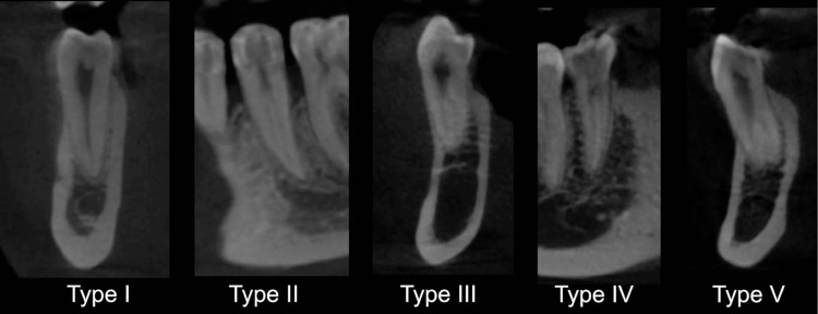
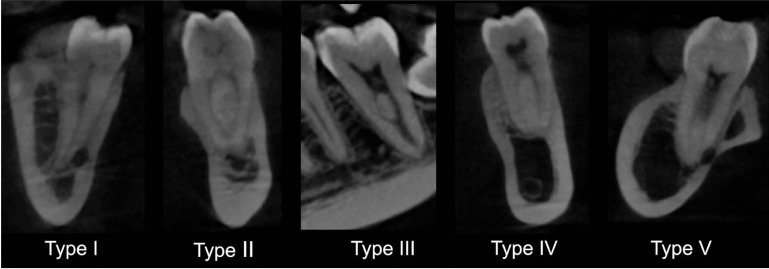
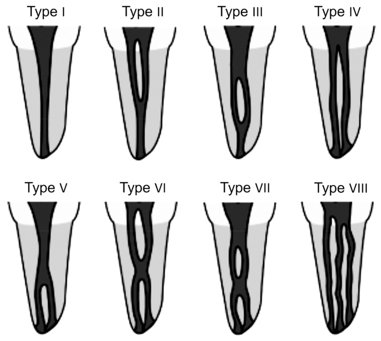
Materials and methods: In total, 121 CBCT images of patients were selected from a database. All images contained lower first and second premolars and molars on both sides of the arch, fully developed roots, and no treatment, resorption, or calcifications. In each image, the root canals of the lower premolars and molars were evaluated according to the Vertucci classification in On-Demand 3D software in the multiplanar reconstruction with dynamic navigation. Twenty-five percent of the images were re-assessed to analyze intraobserver confidence with the kappa test. Data were statistically evaluated with linear regression to evaluate the correlations of anatomic variations with age and sex, and the Wilcoxon test to analyze the laterality of variations, with a significance level of 5%.
Results: The intraobserver agreement (0.94) was excellent. In general, the root canals of lower premolars and molars showed a higher prevalence of type I than other Vertucci classification types, followed by type V in premolars and type II in molars. When the molar roots were evaluated separately, type II was more frequent in mesial roots and type I in distal roots. Although age showed no correlations with the results, sex and laterality showed correlations with tooth 45 and the lower second premolars, respectively.
Conclusion: The lower premolars and molars of a Brazilian sub-population showed a wide range of root canal anatomic variations.

 求助内容:
求助内容: 应助结果提醒方式:


