Solaleh Shahmirzadi, Taraneh Maghsoodi-Zahedi, Sarang Saadat, Husniye Demirturk Kocasarac, Mehrnoosh Rezvan, Rujuta A Katkar, Madhu K Nair
{"title":"Assessment of alveolar bone changes in response to minimally invasive periodontal surgery: A cone-beam computed tomographic evaluation.","authors":"Solaleh Shahmirzadi, Taraneh Maghsoodi-Zahedi, Sarang Saadat, Husniye Demirturk Kocasarac, Mehrnoosh Rezvan, Rujuta A Katkar, Madhu K Nair","doi":"10.5624/isd.20220053","DOIUrl":null,"url":null,"abstract":"<p><strong>Purpose: </strong>The aim of this study was to evaluate 3-dimensional cone-beam computed tomography (CBCT) images of alveolar bone changes in patients who underwent minimally invasive periodontal surgery-namely, the pinhole surgical technique (PST).</p><p><strong>Materials and methods: </strong>Alveolar bone height was measured and compared on CBCT images of 254 teeth from 23 consecutive patients with Miller class I, II, or III recession who had undergone PST. No patient with active periodontal disease was selected for surgery. Two different methods were used to assess the alveolar bone changes postoperatively. In both methods, the distance between the apex of the tooth and the mid-buccal alveolar crestal bone on pre- and post-surgical CBCT studies was measured.</p><p><strong>Results: </strong>An average alveolar bone gain >0.5 mm following PST was identified using CBCT (<i>P</i>=0.05). None of the demographic variables, including sex, age, and time since surgery, had any significant effect on bone gain during follow-up, which ranged from 8 months to 3 years.</p><p><strong>Conclusion: </strong>PST appears to be a promising treatment modality for recession that results in stable clinical outcomes and may lead to some level of resolution on the bone level. More long-term studies must be done to evaluate the impact of this novel technique on bone remodeling and to assess sustained bone levels within a larger study population.</p>","PeriodicalId":51714,"journal":{"name":"Imaging Science in Dentistry","volume":"53 1","pages":"1-9"},"PeriodicalIF":2.1000,"publicationDate":"2023-03-01","publicationTypes":"Journal Article","fieldsOfStudy":null,"isOpenAccess":false,"openAccessPdf":"https://ftp.ncbi.nlm.nih.gov/pub/pmc/oa_pdf/4b/b5/isd-53-1.PMC10060762.pdf","citationCount":"1","resultStr":null,"platform":"Semanticscholar","paperid":null,"PeriodicalName":"Imaging Science in Dentistry","FirstCategoryId":"1085","ListUrlMain":"https://doi.org/10.5624/isd.20220053","RegionNum":0,"RegionCategory":null,"ArticlePicture":[],"TitleCN":null,"AbstractTextCN":null,"PMCID":null,"EPubDate":"","PubModel":"","JCR":"Q3","JCRName":"DENTISTRY, ORAL SURGERY & MEDICINE","Score":null,"Total":0}
引用次数: 1
Abstract
Purpose: The aim of this study was to evaluate 3-dimensional cone-beam computed tomography (CBCT) images of alveolar bone changes in patients who underwent minimally invasive periodontal surgery-namely, the pinhole surgical technique (PST).
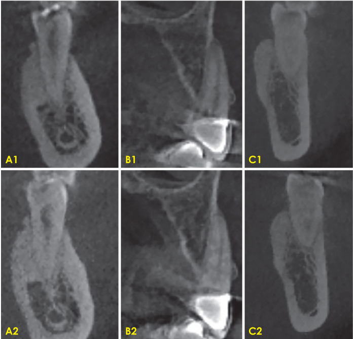
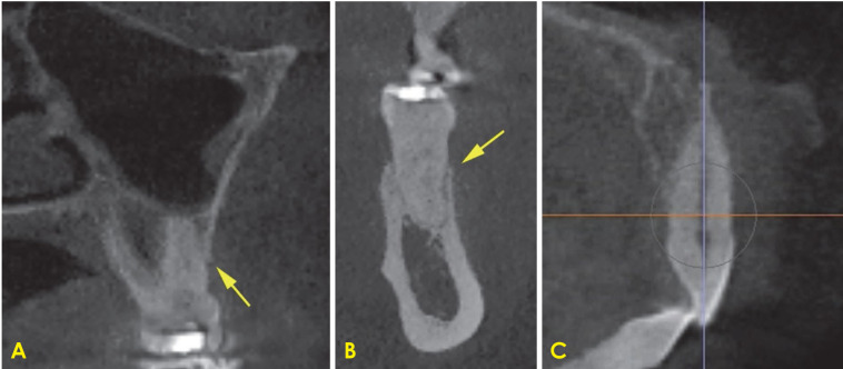
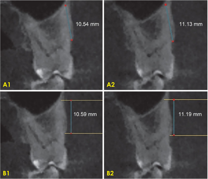
Materials and methods: Alveolar bone height was measured and compared on CBCT images of 254 teeth from 23 consecutive patients with Miller class I, II, or III recession who had undergone PST. No patient with active periodontal disease was selected for surgery. Two different methods were used to assess the alveolar bone changes postoperatively. In both methods, the distance between the apex of the tooth and the mid-buccal alveolar crestal bone on pre- and post-surgical CBCT studies was measured.
Results: An average alveolar bone gain >0.5 mm following PST was identified using CBCT (P=0.05). None of the demographic variables, including sex, age, and time since surgery, had any significant effect on bone gain during follow-up, which ranged from 8 months to 3 years.
Conclusion: PST appears to be a promising treatment modality for recession that results in stable clinical outcomes and may lead to some level of resolution on the bone level. More long-term studies must be done to evaluate the impact of this novel technique on bone remodeling and to assess sustained bone levels within a larger study population.
目的:本研究的目的是评估微创牙周手术(即针孔手术技术(PST))患者的牙槽骨变化的三维锥形束计算机断层扫描(CBCT)图像。材料和方法:对23例连续行PST的Miller I、II、III级衰退患者的254颗牙的CBCT图像进行牙槽骨高度测量和比较。没有选择活动性牙周病患者进行手术。采用两种不同的方法评估术后牙槽骨的变化。在这两种方法中,在术前和术后的CBCT研究中测量了牙尖与中颊牙槽嵴骨之间的距离。结果:CBCT显示PST术后牙槽骨平均增厚>0.5 mm (P=0.05)。在随访期间(8个月至3年),包括性别、年龄和手术后时间在内的人口统计学变量均未对骨增重产生显著影响。结论:PST似乎是一种有希望的治疗衰退的方式,结果稳定的临床结果,并可能导致一定程度的骨水平的解决。必须进行更多的长期研究来评估这种新技术对骨重塑的影响,并在更大的研究人群中评估持续的骨水平。

 求助内容:
求助内容: 应助结果提醒方式:


