{"title":"Assessment of Radiodensity at Mandibular Periapical Bone Sites using Three-Dimensional Cone-Beam Computed Tomography.","authors":"Samir Goyushov, Neset Volkan Asar, Tolga Fikret Tözüm","doi":"10.5037/jomr.2023.14102","DOIUrl":null,"url":null,"abstract":"<p><strong>Objectives: </strong>The aims of this retrospective study were to objectively assess bone density values obtained by cone-beam computed tomography and to map the periapical and inter-radicular regions of the mandibular bone.</p><p><strong>Material and methods: </strong>In total, periapical bone regions of 6898 roots scanned by cone-beam computed tomography were evaluated retrospectively, and the results were recorded using Hounsfield units (HU).</p><p><strong>Results: </strong>The correlation between periapical HU values of adjacent mandibular teeth were strongly positive (P ˂ 0.01). The anterior region of the mandible yielded highest mean HU value (633.55). The mean periapical HU value of the premolar region (470.58) was higher than that was measured for molar region (374.58). The difference between furcation HU values of the first and second molars was unnoticeable.</p><p><strong>Conclusions: </strong>The results of this study have tried to evaluate the periapical regions of all mandibular teeth, which could ease to predict the bone radiodensity before implant surgery. Even though the Hounsfield units provide the average radio-bone density, a site-specific bone tissue evaluation of each case is essential for appropriate cone-beam computed tomography preoperative planning.</p>","PeriodicalId":53254,"journal":{"name":"eJournal of Oral Maxillofacial Research","volume":"14 1","pages":"e2"},"PeriodicalIF":1.0000,"publicationDate":"2023-01-01","publicationTypes":"Journal Article","fieldsOfStudy":null,"isOpenAccess":false,"openAccessPdf":"https://ftp.ncbi.nlm.nih.gov/pub/pmc/oa_pdf/39/17/jomr-14-e2.PMC10170661.pdf","citationCount":"1","resultStr":null,"platform":"Semanticscholar","paperid":null,"PeriodicalName":"eJournal of Oral Maxillofacial Research","FirstCategoryId":"1085","ListUrlMain":"https://doi.org/10.5037/jomr.2023.14102","RegionNum":0,"RegionCategory":null,"ArticlePicture":[],"TitleCN":null,"AbstractTextCN":null,"PMCID":null,"EPubDate":"","PubModel":"","JCR":"Q3","JCRName":"DENTISTRY, ORAL SURGERY & MEDICINE","Score":null,"Total":0}
引用次数: 1
Abstract
Objectives: The aims of this retrospective study were to objectively assess bone density values obtained by cone-beam computed tomography and to map the periapical and inter-radicular regions of the mandibular bone.
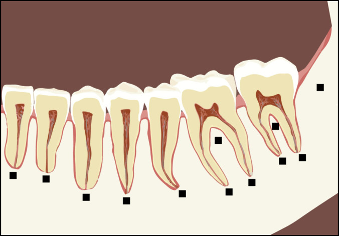
Material and methods: In total, periapical bone regions of 6898 roots scanned by cone-beam computed tomography were evaluated retrospectively, and the results were recorded using Hounsfield units (HU).
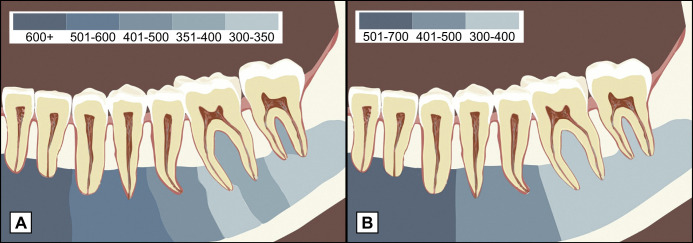
Results: The correlation between periapical HU values of adjacent mandibular teeth were strongly positive (P ˂ 0.01). The anterior region of the mandible yielded highest mean HU value (633.55). The mean periapical HU value of the premolar region (470.58) was higher than that was measured for molar region (374.58). The difference between furcation HU values of the first and second molars was unnoticeable.
Conclusions: The results of this study have tried to evaluate the periapical regions of all mandibular teeth, which could ease to predict the bone radiodensity before implant surgery. Even though the Hounsfield units provide the average radio-bone density, a site-specific bone tissue evaluation of each case is essential for appropriate cone-beam computed tomography preoperative planning.

 求助内容:
求助内容: 应助结果提醒方式:


