{"title":"Clinico-cytomorphological Spectrum of Calcinosis Cutis.","authors":"Malvika Shastri, Pratibha Gautam, Preeti Diwaker, Priyanka Gogoi, Vinod K Arora","doi":"10.4103/joc.joc_75_22","DOIUrl":null,"url":null,"abstract":"<p><strong>Introduction: </strong>The deposition of calcium in the skin is known as calcinosis cutis. It can affect any part of the body and can mimic soft tissue or bony lesions clinically.</p><p><strong>Aim: </strong>To describe the clinical and cytomorphologic features of calcinosis cutis on fine needle aspiration cytology smears.</p><p><strong>Materials and methods: </strong>A total of 17 cases reported as calcinosis cutis on fine needle aspiration cytology were reviewed for the available clinical and cytological details.</p><p><strong>Results: </strong>The cohort included both adult and pediatric patients. Clinically, the lesions appeared as painless swellings of variable sizes. The common sites affected were the scrotum, iliac region, scalp, pinna, neck, axilla, elbow, arm, thigh, and gluteal region. Aspirate was chalky white, paste-like in all the cases. The cytologic evaluation revealed amorphous crystalline deposits of calcium along with histiocytes, lymphocytes, and multinucleated giant cells.</p><p><strong>Conclusions: </strong>Calcinosis cutis has a wide spectrum of clinical presentations. Fine needle aspiration cytology is a minimally invasive approach for diagnosing calcinosis cutis, thus eliminating the need for more extensive biopsy procedures.</p>","PeriodicalId":50217,"journal":{"name":"Journal of Cytology","volume":"40 1","pages":"24-27"},"PeriodicalIF":1.0000,"publicationDate":"2023-01-01","publicationTypes":"Journal Article","fieldsOfStudy":null,"isOpenAccess":false,"openAccessPdf":"https://www.ncbi.nlm.nih.gov/pmc/articles/PMC10167833/pdf/","citationCount":"0","resultStr":null,"platform":"Semanticscholar","paperid":null,"PeriodicalName":"Journal of Cytology","FirstCategoryId":"3","ListUrlMain":"https://doi.org/10.4103/joc.joc_75_22","RegionNum":4,"RegionCategory":"医学","ArticlePicture":[],"TitleCN":null,"AbstractTextCN":null,"PMCID":null,"EPubDate":"2023/3/13 0:00:00","PubModel":"Epub","JCR":"Q4","JCRName":"MEDICAL LABORATORY TECHNOLOGY","Score":null,"Total":0}
引用次数: 0
Abstract
Introduction: The deposition of calcium in the skin is known as calcinosis cutis. It can affect any part of the body and can mimic soft tissue or bony lesions clinically.
Aim: To describe the clinical and cytomorphologic features of calcinosis cutis on fine needle aspiration cytology smears.
Materials and methods: A total of 17 cases reported as calcinosis cutis on fine needle aspiration cytology were reviewed for the available clinical and cytological details.
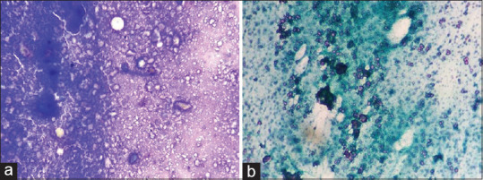
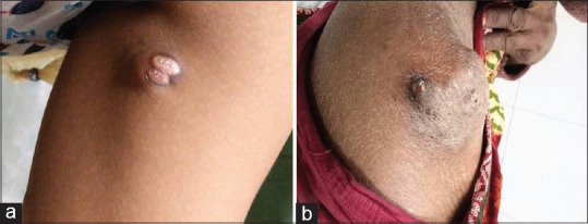
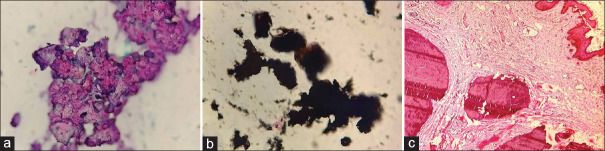
Results: The cohort included both adult and pediatric patients. Clinically, the lesions appeared as painless swellings of variable sizes. The common sites affected were the scrotum, iliac region, scalp, pinna, neck, axilla, elbow, arm, thigh, and gluteal region. Aspirate was chalky white, paste-like in all the cases. The cytologic evaluation revealed amorphous crystalline deposits of calcium along with histiocytes, lymphocytes, and multinucleated giant cells.
Conclusions: Calcinosis cutis has a wide spectrum of clinical presentations. Fine needle aspiration cytology is a minimally invasive approach for diagnosing calcinosis cutis, thus eliminating the need for more extensive biopsy procedures.
期刊介绍:
The Journal of Cytology is the official Quarterly publication of the Indian Academy of Cytologists. It is in the 25th year of publication in the year 2008. The journal covers all aspects of diagnostic cytology, including fine needle aspiration cytology, gynecological and non-gynecological cytology. Articles on ancillary techniques, like cytochemistry, immunocytochemistry, electron microscopy, molecular cytopathology, as applied to cytological material are also welcome. The journal gives preference to clinically oriented studies over experimental and animal studies. The Journal would publish peer-reviewed original research papers, case reports, systematic reviews, meta-analysis, and debates.
 求助内容:
求助内容: 应助结果提醒方式:


