{"title":"Extraabdominal parasitic lipoleiomyoma.","authors":"Tae Hoon Lee, Se-Jin Baek","doi":"10.14216/kjco.21008","DOIUrl":null,"url":null,"abstract":"<p><p>Extrauterine parasitic lipoleiomyoma is a very rare fatty tumor, with uncertain histopathogenesis. Although imaging studies play an important role in preoperative localization and diagnosis of lipoleiomyoma, a pathological evaluation is paramount for confirmation of diagnosis. We describe a case of a 49-year-old woman with a palpable mass in the right inguinal area. Computed tomography of the abdomen and pelvis revealed a fluid- and fat-containing mass. Histopathological examination of the mass, which was successfully resected, confirmed the diagnosis of lipoleiomyoma. The patient was discharged on a postoperative day 2 without any complications.</p>","PeriodicalId":74045,"journal":{"name":"Korean journal of clinical oncology","volume":"17 1","pages":"48-51"},"PeriodicalIF":0.0000,"publicationDate":"2021-06-01","publicationTypes":"Journal Article","fieldsOfStudy":null,"isOpenAccess":false,"openAccessPdf":"https://ftp.ncbi.nlm.nih.gov/pub/pmc/oa_pdf/83/50/kjco-17-1-48.PMC9942745.pdf","citationCount":"1","resultStr":null,"platform":"Semanticscholar","paperid":null,"PeriodicalName":"Korean journal of clinical oncology","FirstCategoryId":"1085","ListUrlMain":"https://doi.org/10.14216/kjco.21008","RegionNum":0,"RegionCategory":null,"ArticlePicture":[],"TitleCN":null,"AbstractTextCN":null,"PMCID":null,"EPubDate":"","PubModel":"","JCR":"","JCRName":"","Score":null,"Total":0}
引用次数: 1
Abstract
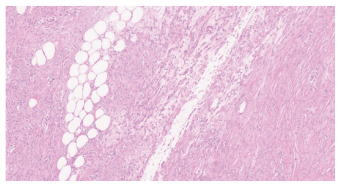
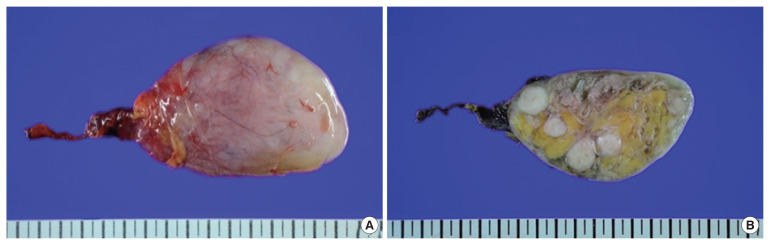
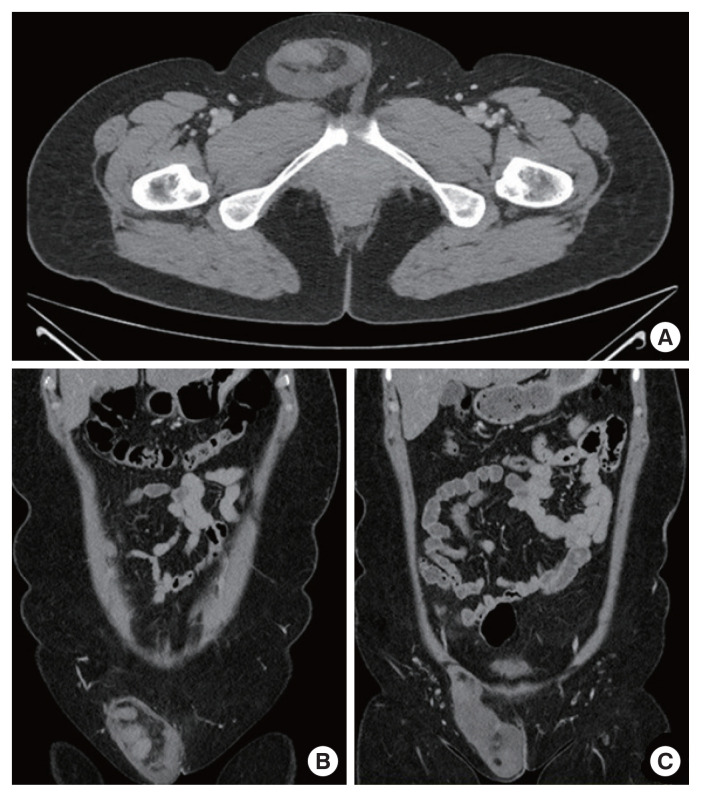
Extrauterine parasitic lipoleiomyoma is a very rare fatty tumor, with uncertain histopathogenesis. Although imaging studies play an important role in preoperative localization and diagnosis of lipoleiomyoma, a pathological evaluation is paramount for confirmation of diagnosis. We describe a case of a 49-year-old woman with a palpable mass in the right inguinal area. Computed tomography of the abdomen and pelvis revealed a fluid- and fat-containing mass. Histopathological examination of the mass, which was successfully resected, confirmed the diagnosis of lipoleiomyoma. The patient was discharged on a postoperative day 2 without any complications.

 求助内容:
求助内容: 应助结果提醒方式:


