Zhigang Rong, Zhong Yang, Chengmin Zhang, Rongxi Pu, Can Chen, Jianzhong Xu, Fei Luo
{"title":"Bioinformatics analysis of paravertebral muscles atrophy in adult degenerative scoliosis.","authors":"Zhigang Rong, Zhong Yang, Chengmin Zhang, Rongxi Pu, Can Chen, Jianzhong Xu, Fei Luo","doi":"10.1007/s10974-023-09650-8","DOIUrl":null,"url":null,"abstract":"<p><p>Paravertebral muscles (PVM) act as one of the major dynamic factors to maintain human upright activities and play a remarkable role in maintaining the balance of the trunk. Adult degenerative scoliosis (ADS) has become one of the important causes of disability in the elderly population owing to the changes in spinal biomechanics, atrophy and degeneration of PVM, and imbalance of the spine. Previously, many studies focused on the physical evaluation of PVM degeneration. However, the molecular biological changes are still not completely known. In this study, we established a rat model of scoliosis and performed the proteomic analysis of the PVM of ADS. The results showed that the degree of atrophy, muscle fat deposition, and fibrosis of the PVM of rats positively correlated with the angle of scoliosis. The proteomic results showed that 177 differentially expressed proteins were present in the ADS group, which included 105 upregulated proteins and 72 downregulated proteins compared with the PVM in individuals without spinal deformities. Through the construction of a protein-protein interaction network, 18 core differentially expressed proteins were obtained, which included fibrinogen beta chain, apolipoprotein E, fibrinogen gamma chain, thrombospondin-1, integrin alpha-6, fibronectin-1, platelet factor 4, coagulation factor XIII A chain, ras-related protein Rap-1b, platelet endothelial cell adhesion molecule 1, complement C1q subcomponent subunit A, cathepsin G, myeloperoxidase, von Willebrand factor, integrin beta-1, integrin alpha-1, leukocyte surface antigen CD47, and complement C1q subcomponent subunit B. Further analysis of the Kyoto Encyclopedia of Genes and Genomes pathway (KEGG) and immunofluorescence showed that the neutrophil extracellular traps (NETs) formation signaling pathway plays a major role in the pathogenesis of PVM degeneration in ADS. The results of the present study preliminarily laid the molecular biological foundation of PVM atrophy in ADS, which will provide a new therapeutic target for alleviating PVM atrophy and decreasing the occurrence of scoliosis.</p>","PeriodicalId":16422,"journal":{"name":"Journal of Muscle Research and Cell Motility","volume":" ","pages":"287-297"},"PeriodicalIF":1.7000,"publicationDate":"2023-12-01","publicationTypes":"Journal Article","fieldsOfStudy":null,"isOpenAccess":false,"openAccessPdf":"https://www.ncbi.nlm.nih.gov/pmc/articles/PMC10665243/pdf/","citationCount":"0","resultStr":null,"platform":"Semanticscholar","paperid":null,"PeriodicalName":"Journal of Muscle Research and Cell Motility","FirstCategoryId":"99","ListUrlMain":"https://doi.org/10.1007/s10974-023-09650-8","RegionNum":3,"RegionCategory":"生物学","ArticlePicture":[],"TitleCN":null,"AbstractTextCN":null,"PMCID":null,"EPubDate":"2023/5/20 0:00:00","PubModel":"Epub","JCR":"Q4","JCRName":"CELL BIOLOGY","Score":null,"Total":0}
引用次数: 0
Abstract
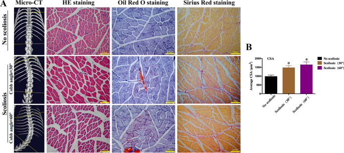
Paravertebral muscles (PVM) act as one of the major dynamic factors to maintain human upright activities and play a remarkable role in maintaining the balance of the trunk. Adult degenerative scoliosis (ADS) has become one of the important causes of disability in the elderly population owing to the changes in spinal biomechanics, atrophy and degeneration of PVM, and imbalance of the spine. Previously, many studies focused on the physical evaluation of PVM degeneration. However, the molecular biological changes are still not completely known. In this study, we established a rat model of scoliosis and performed the proteomic analysis of the PVM of ADS. The results showed that the degree of atrophy, muscle fat deposition, and fibrosis of the PVM of rats positively correlated with the angle of scoliosis. The proteomic results showed that 177 differentially expressed proteins were present in the ADS group, which included 105 upregulated proteins and 72 downregulated proteins compared with the PVM in individuals without spinal deformities. Through the construction of a protein-protein interaction network, 18 core differentially expressed proteins were obtained, which included fibrinogen beta chain, apolipoprotein E, fibrinogen gamma chain, thrombospondin-1, integrin alpha-6, fibronectin-1, platelet factor 4, coagulation factor XIII A chain, ras-related protein Rap-1b, platelet endothelial cell adhesion molecule 1, complement C1q subcomponent subunit A, cathepsin G, myeloperoxidase, von Willebrand factor, integrin beta-1, integrin alpha-1, leukocyte surface antigen CD47, and complement C1q subcomponent subunit B. Further analysis of the Kyoto Encyclopedia of Genes and Genomes pathway (KEGG) and immunofluorescence showed that the neutrophil extracellular traps (NETs) formation signaling pathway plays a major role in the pathogenesis of PVM degeneration in ADS. The results of the present study preliminarily laid the molecular biological foundation of PVM atrophy in ADS, which will provide a new therapeutic target for alleviating PVM atrophy and decreasing the occurrence of scoliosis.
期刊介绍:
The Journal of Muscle Research and Cell Motility has as its main aim the publication of original research which bears on either the excitation and contraction of muscle, the analysis of any one of the processes involved therein, the processes underlying contractility and motility of animal and plant cells, the toxicology and pharmacology related to contractility, or the formation, dynamics and turnover of contractile structures in muscle and non-muscle cells. Studies describing the impact of pathogenic mutations in genes encoding components of contractile structures in humans or animals are welcome, provided they offer mechanistic insight into the disease process or the underlying gene function. The policy of the Journal is to encourage any form of novel practical study whatever its specialist interest, as long as it falls within this broad field. Theoretical essays are welcome provided that they are concise and suggest practical ways in which they may be tested. Manuscripts reporting new mutations in known disease genes without validation and mechanistic insight will not be considered. It is the policy of the journal that cells lines, hybridomas and DNA clones should be made available by the developers to any qualified investigator. Submission of a manuscript for publication constitutes an agreement of the authors to abide by this principle.
 求助内容:
求助内容: 应助结果提醒方式:


