Doris Lambracht-Washington , Min Fu , Navid Manouchehri , Linda S. Hynan , Olaf Stuve , Roger N. Rosenberg
{"title":"Glial cell transcriptome analyses in 3xTg-AD mice: Effects of aging, disease progression, and anti-Aβ immunotherapy","authors":"Doris Lambracht-Washington , Min Fu , Navid Manouchehri , Linda S. Hynan , Olaf Stuve , Roger N. Rosenberg","doi":"10.1016/j.nbas.2023.100066","DOIUrl":null,"url":null,"abstract":"<div><h3>Background</h3><p>To investigate how changes in expression of glial genes relate to a progression of Alzheimer’s disease (AD) pathology, and how anti-Aβ immunotherapy impact these changes, we conducted a transcriptomic analysis for brains from cohorts of 2-, 10-, and 20 month old 3xTg-AD mice, and a cross-sectional study in groups of 20 month-old mice treated with active DNA Aβ42 immunization, passive immunotherapy, untreated, and wild-type (wt) controls.</p></div><div><h3>Methods</h3><p>Twenty-four Formalin-Fixed Paraffin-Embedded (FFPE) mouse brain sections were used for the gene expression analyses (nanostring). Adjacent sections from these and additional mouse brains were stained for microglia using antibodies detecting IbaI and Gal3. For a semi-quantitative analysis of increased tau and amyloid pathology with aging and disease progression, a comparison of ELISA results from brains of 12 and 20 months old 3xTg-AD mice were shown.</p></div><div><h3>Results</h3><p>Based on the different comparisons of transcript numbers found the 3xTg-AD age groups with the senescent 20 months old wt control mouse brains, and the 20 months old 3xTg-AD mouse brains with the 20 months old wt control mouse brains, genes were assigned as upregulated due to aging, or due to disease progression, or due to both. The immunohistochemistry of microglia markers revealed that Gal3 might be an important marker for phagocytosing microglia around amyloid plaques. The comparison of the two anti-Aβ immunotherapy approaches showed a differential downregulation of inflammatory glial genes.</p></div><div><h3>Conclusion</h3><p>These results are relevant for future clinical trials using active anti-amyloid immunotherapy.</p></div>","PeriodicalId":72131,"journal":{"name":"Aging brain","volume":"3 ","pages":"Article 100066"},"PeriodicalIF":1.7000,"publicationDate":"2023-01-01","publicationTypes":"Journal Article","fieldsOfStudy":null,"isOpenAccess":false,"openAccessPdf":"https://ftp.ncbi.nlm.nih.gov/pub/pmc/oa_pdf/f3/6f/main.PMC9997156.pdf","citationCount":"0","resultStr":null,"platform":"Semanticscholar","paperid":null,"PeriodicalName":"Aging brain","FirstCategoryId":"1085","ListUrlMain":"https://www.sciencedirect.com/science/article/pii/S2589958923000038","RegionNum":0,"RegionCategory":null,"ArticlePicture":[],"TitleCN":null,"AbstractTextCN":null,"PMCID":null,"EPubDate":"","PubModel":"","JCR":"Q3","JCRName":"CLINICAL NEUROLOGY","Score":null,"Total":0}
引用次数: 0
Abstract
Background
To investigate how changes in expression of glial genes relate to a progression of Alzheimer’s disease (AD) pathology, and how anti-Aβ immunotherapy impact these changes, we conducted a transcriptomic analysis for brains from cohorts of 2-, 10-, and 20 month old 3xTg-AD mice, and a cross-sectional study in groups of 20 month-old mice treated with active DNA Aβ42 immunization, passive immunotherapy, untreated, and wild-type (wt) controls.
Methods
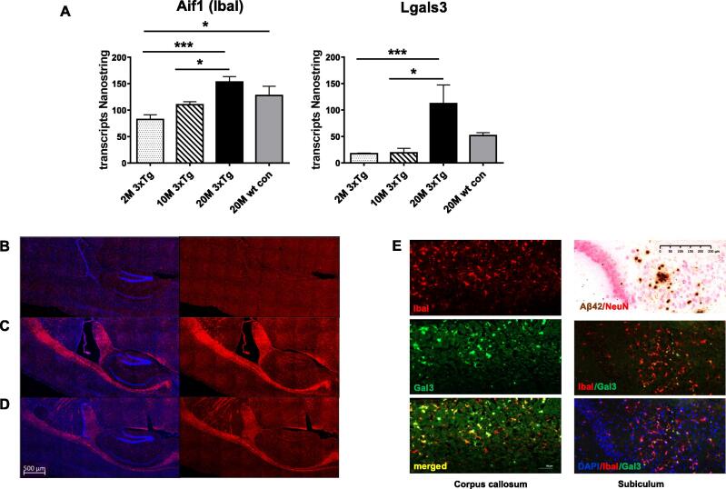
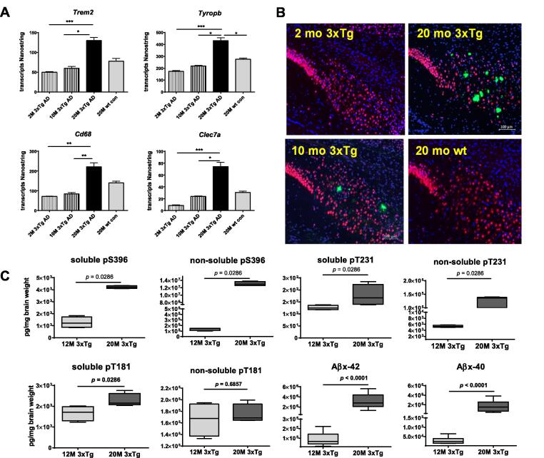
Twenty-four Formalin-Fixed Paraffin-Embedded (FFPE) mouse brain sections were used for the gene expression analyses (nanostring). Adjacent sections from these and additional mouse brains were stained for microglia using antibodies detecting IbaI and Gal3. For a semi-quantitative analysis of increased tau and amyloid pathology with aging and disease progression, a comparison of ELISA results from brains of 12 and 20 months old 3xTg-AD mice were shown.
Results
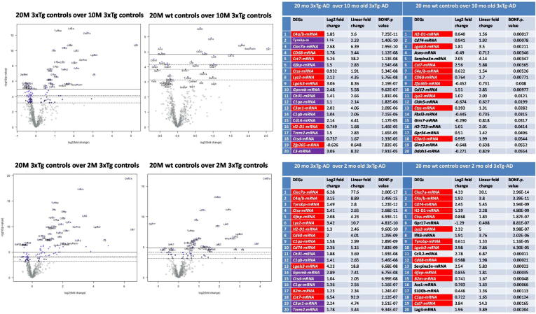
Based on the different comparisons of transcript numbers found the 3xTg-AD age groups with the senescent 20 months old wt control mouse brains, and the 20 months old 3xTg-AD mouse brains with the 20 months old wt control mouse brains, genes were assigned as upregulated due to aging, or due to disease progression, or due to both. The immunohistochemistry of microglia markers revealed that Gal3 might be an important marker for phagocytosing microglia around amyloid plaques. The comparison of the two anti-Aβ immunotherapy approaches showed a differential downregulation of inflammatory glial genes.
Conclusion
These results are relevant for future clinical trials using active anti-amyloid immunotherapy.
为了研究神经胶质基因表达的变化与阿尔茨海默病(AD)病理进展的关系,以及抗β免疫治疗如何影响这些变化,我们对2、10和20月龄3xTg-AD小鼠的大脑进行了转录组学分析,并对20月龄小鼠进行了横断面研究,这些小鼠分别接受了主动DNA a β42免疫、被动免疫治疗、未治疗和野生型(wt)对照。方法采用24张福尔马林固定石蜡包埋(FFPE)小鼠脑切片进行基因表达分析(纳米链)。使用检测IbaI和Gal3的抗体对这些和其他小鼠大脑的邻近切片进行小胶质细胞染色。为了半定量分析随着衰老和疾病进展而增加的tau和淀粉样蛋白病理,对12个月和20个月大3xTg-AD小鼠大脑的ELISA结果进行了比较。结果通过对3xTg-AD年龄组与衰老的20月龄wt对照小鼠脑,以及20月龄3xTg-AD小鼠脑与20月龄wt对照小鼠脑的转录本数量的不同比较发现,基因被定位为由于衰老或由于疾病进展而上调,或由于两者兼有。小胶质细胞标记物免疫组化结果显示,Gal3可能是吞噬淀粉样斑块周围小胶质细胞的重要标记物。两种抗a β免疫治疗方法的比较显示炎症胶质基因的差异下调。结论本研究结果对今后应用主动抗淀粉样蛋白免疫疗法进行临床试验具有一定的指导意义。

 求助内容:
求助内容: 应助结果提醒方式:


