{"title":"Is Type 2 Macular Telangiectasia a Bilateral and Symmetrical Disease Entity?","authors":"Ramesh Venkatesh, Harshita Nahata, Nikitha Gurram Reddy, Pranjal Mishra, Rubble Mangla, Naresh Kumar Yadav, Jay Chhablani","doi":"10.4103/joco.joco_68_22","DOIUrl":null,"url":null,"abstract":"<p><strong>Purpose: </strong>To study the inter-eye asymmetry in cases diagnosed with type 2 macular telangiectasia (MacTel).</p><p><strong>Methods: </strong>Herein, type 2 MacTel cases were staged as per Gass and Blodi classification with multiple imaging techniques. Based on disease stage symmetry, two groups identified. Group 1: Symmetrical stage and Group 2: Asymmetrical stage MacTel disease. Prevalence, demography, and clinical features of MacTel cases showing inter-eye asymmetry were analyzed.</p><p><strong>Results: </strong>Two hundred and eighty eyes of 140 patients diagnosed clinically with type 2 MacTel (84-Group 1 and 56-Group 2) were evaluated. Eighty-nine (64%) were female, and the median age of the entire cohort was 62.5 years (inter-quartile range: 57.0-68.75). MacTel disease with asymmetric stage was seen in 56 (40%) of the 140 patients. At presentation, a two-stage difference was noted in 46% (<i>n</i> = 26) of the patients with asymmetrical MacTel disease. A 10% conversion from symmetrical to asymmetrical disease stage was noted at the final visit. Of the 280 eyes evaluated for type 2 MacTel disease, 12 (4%) eyes showed no findings suggestive of MacTel on clinical examination and fluorescein angiography, optical coherence tomography (OCT), and OCT angiography when available and were labeled as unilateral type 2 MacTel disease.</p><p><strong>Conclusions: </strong>Type 2 MacTel can show inter-eye disease stage asymmetry. Unilateral type 2 MacTel disease is a distinct stage in MacTel which would need further evaluation and consideration while staging.</p>","PeriodicalId":15423,"journal":{"name":"Journal of Current Ophthalmology","volume":null,"pages":null},"PeriodicalIF":1.2000,"publicationDate":"2022-10-01","publicationTypes":"Journal Article","fieldsOfStudy":null,"isOpenAccess":false,"openAccessPdf":"https://ftp.ncbi.nlm.nih.gov/pub/pmc/oa_pdf/b5/7f/JCO-34-428.PMC10170975.pdf","citationCount":"1","resultStr":null,"platform":"Semanticscholar","paperid":null,"PeriodicalName":"Journal of Current Ophthalmology","FirstCategoryId":"1085","ListUrlMain":"https://doi.org/10.4103/joco.joco_68_22","RegionNum":0,"RegionCategory":null,"ArticlePicture":[],"TitleCN":null,"AbstractTextCN":null,"PMCID":null,"EPubDate":"","PubModel":"","JCR":"Q3","JCRName":"OPHTHALMOLOGY","Score":null,"Total":0}
引用次数: 1
Abstract
Purpose: To study the inter-eye asymmetry in cases diagnosed with type 2 macular telangiectasia (MacTel).
Methods: Herein, type 2 MacTel cases were staged as per Gass and Blodi classification with multiple imaging techniques. Based on disease stage symmetry, two groups identified. Group 1: Symmetrical stage and Group 2: Asymmetrical stage MacTel disease. Prevalence, demography, and clinical features of MacTel cases showing inter-eye asymmetry were analyzed.
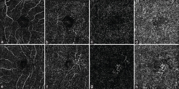
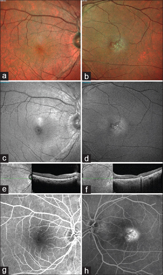
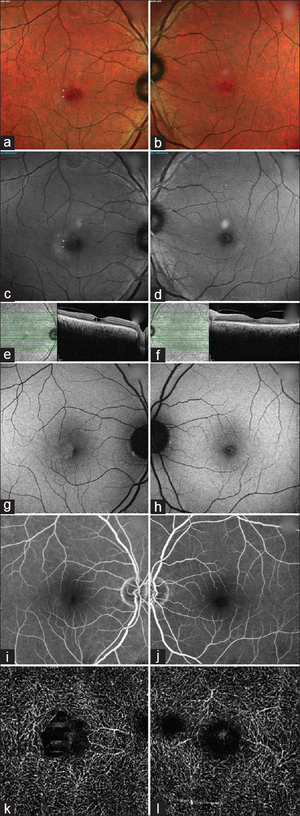
Results: Two hundred and eighty eyes of 140 patients diagnosed clinically with type 2 MacTel (84-Group 1 and 56-Group 2) were evaluated. Eighty-nine (64%) were female, and the median age of the entire cohort was 62.5 years (inter-quartile range: 57.0-68.75). MacTel disease with asymmetric stage was seen in 56 (40%) of the 140 patients. At presentation, a two-stage difference was noted in 46% (n = 26) of the patients with asymmetrical MacTel disease. A 10% conversion from symmetrical to asymmetrical disease stage was noted at the final visit. Of the 280 eyes evaluated for type 2 MacTel disease, 12 (4%) eyes showed no findings suggestive of MacTel on clinical examination and fluorescein angiography, optical coherence tomography (OCT), and OCT angiography when available and were labeled as unilateral type 2 MacTel disease.
Conclusions: Type 2 MacTel can show inter-eye disease stage asymmetry. Unilateral type 2 MacTel disease is a distinct stage in MacTel which would need further evaluation and consideration while staging.
期刊介绍:
Peer Review under the responsibility of Iranian Society of Ophthalmology Journal of Current Ophthalmology, the official publication of the Iranian Society of Ophthalmology, is a peer-reviewed, open-access, scientific journal that welcomes high quality original articles related to vision science and all fields of ophthalmology. Journal of Current Ophthalmology is the continuum of Iranian Journal of Ophthalmology published since 1969.

 求助内容:
求助内容: 应助结果提醒方式:


