Mohammad Yaser Kiarudi, Bahar Tafaghodi, Aliakbar Sabermoghadm, Acieh Es'haghi, Seyed Hosein Ghavami Shahri
{"title":"Medial Rectus Plication in the Management of Dissociated Horizontal Deviation: Case Report and Literature Review.","authors":"Mohammad Yaser Kiarudi, Bahar Tafaghodi, Aliakbar Sabermoghadm, Acieh Es'haghi, Seyed Hosein Ghavami Shahri","doi":"10.4103/joco.joco_6_22","DOIUrl":null,"url":null,"abstract":"<p><strong>Purpose: </strong>To report a case of medial rectus plication for the management of dissociated horizontal deviation (DHD).</p><p><strong>Methods: </strong>We introduce medial rectus plication for improving the control of exoshift of DHD.</p><p><strong>Results: </strong>A 20-year-old woman with a chief complaint of left eye outward deviation since childhood was referred to the strabismus clinic. The diagnosis of DHD was made according to the detection of asymmetric slow abduction of the left eye (50 prism diopter) during visual inattention or cover testing. The left lateral rectus (LR) was recessed 8 mm with a posterior fixation suture (PFS). In the early postoperative period, the control of DHD improved; however, after 6 months, the patient and her parents complained of frequent observation of the exoshift of the left eye (30 prism diopter). For better control of DHD, medial rectus plication (5 mm) of the left eye was considered the second operation. After 12 months of follow-up, the control of deviation improved, and there was no manifest deviation.</p><p><strong>Conclusions: </strong>The literature's recommended procedure for unilateral DHD without a duction deficit is to perform a unilateral LR muscle recession. Some authors have proposed adding PFS to augment the effect of LR recessions. Although recurrence may occur, medial rectus plication can be considered one of the reversible options and can be used in recurrences of DHD after the first surgical procedure.</p>","PeriodicalId":15423,"journal":{"name":"Journal of Current Ophthalmology","volume":null,"pages":null},"PeriodicalIF":1.2000,"publicationDate":"2022-10-01","publicationTypes":"Journal Article","fieldsOfStudy":null,"isOpenAccess":false,"openAccessPdf":"https://ftp.ncbi.nlm.nih.gov/pub/pmc/oa_pdf/c5/3e/JCO-34-483.PMC10170982.pdf","citationCount":"0","resultStr":null,"platform":"Semanticscholar","paperid":null,"PeriodicalName":"Journal of Current Ophthalmology","FirstCategoryId":"1085","ListUrlMain":"https://doi.org/10.4103/joco.joco_6_22","RegionNum":0,"RegionCategory":null,"ArticlePicture":[],"TitleCN":null,"AbstractTextCN":null,"PMCID":null,"EPubDate":"","PubModel":"","JCR":"Q3","JCRName":"OPHTHALMOLOGY","Score":null,"Total":0}
引用次数: 0
Abstract
Purpose: To report a case of medial rectus plication for the management of dissociated horizontal deviation (DHD).
Methods: We introduce medial rectus plication for improving the control of exoshift of DHD.
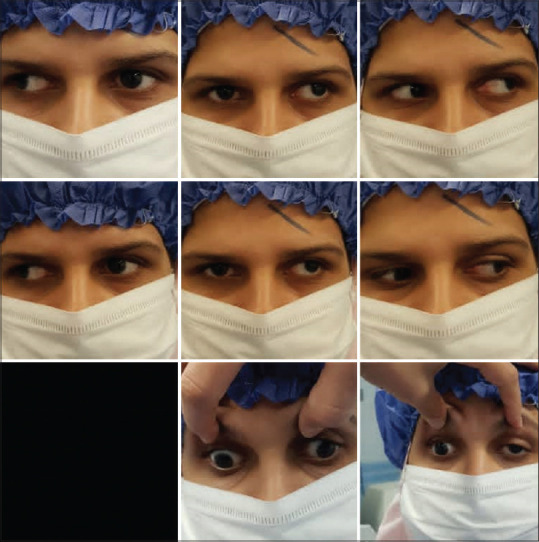
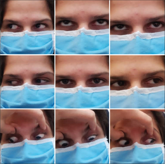
Results: A 20-year-old woman with a chief complaint of left eye outward deviation since childhood was referred to the strabismus clinic. The diagnosis of DHD was made according to the detection of asymmetric slow abduction of the left eye (50 prism diopter) during visual inattention or cover testing. The left lateral rectus (LR) was recessed 8 mm with a posterior fixation suture (PFS). In the early postoperative period, the control of DHD improved; however, after 6 months, the patient and her parents complained of frequent observation of the exoshift of the left eye (30 prism diopter). For better control of DHD, medial rectus plication (5 mm) of the left eye was considered the second operation. After 12 months of follow-up, the control of deviation improved, and there was no manifest deviation.
Conclusions: The literature's recommended procedure for unilateral DHD without a duction deficit is to perform a unilateral LR muscle recession. Some authors have proposed adding PFS to augment the effect of LR recessions. Although recurrence may occur, medial rectus plication can be considered one of the reversible options and can be used in recurrences of DHD after the first surgical procedure.
期刊介绍:
Peer Review under the responsibility of Iranian Society of Ophthalmology Journal of Current Ophthalmology, the official publication of the Iranian Society of Ophthalmology, is a peer-reviewed, open-access, scientific journal that welcomes high quality original articles related to vision science and all fields of ophthalmology. Journal of Current Ophthalmology is the continuum of Iranian Journal of Ophthalmology published since 1969.

 求助内容:
求助内容: 应助结果提醒方式:


