Cassidy Anderson, Aishwarya Sriram, Abigail Funari, Kevin Hsu, Raquel Yokoda, Isabella Pecorari, Isabella Flaquer, Nadeem Akbar, Patrick Colley, Geoffrey Basson, Howard S Moskowitz, Vijay Agarwal
{"title":"High-Grade Ectopic Pituitary Adenoma within the Cerebellopontine Angle: A Case Report.","authors":"Cassidy Anderson, Aishwarya Sriram, Abigail Funari, Kevin Hsu, Raquel Yokoda, Isabella Pecorari, Isabella Flaquer, Nadeem Akbar, Patrick Colley, Geoffrey Basson, Howard S Moskowitz, Vijay Agarwal","doi":"10.1055/a-2065-9809","DOIUrl":null,"url":null,"abstract":"<p><p>Ectopic pituitary tumors are neoplasms with no connection to the pituitary gland and are commonly deposited in other areas of the anterior skull base. A 32-year-old woman presented with a 3-month history of right-sided facial weakness, sensorineural hearing loss, diplopia, and severe headaches. Physical examination revealed a mid-dilated sluggishly reactive right pupil with slight limitation in all gazes, as well as right-sided orbicularis weakness, lagophthalmos, and decreased facial sensation. A magnetic resonance imaging (MRI) of the head without contrast revealed a 3.7 × 1.8 × 2.6 cm mildly enhancing mass in the right internal acoustic meatus and along the petrous ridge. The case was brought before the institution's tumor board, where concern for higher grade pathology, such as hemangiopericytoma, was discussed. Per patient preference, surgical biopsy of the tumor was performed. Immunohistochemical staining revealed a World Health Organization (WHO) grade II neuroendocrine tumor, with cells staining positive for synaptophysin, chromogranin, and CD56, with a K <sub>i</sub> -67 index of 8%. In addition to the ectopic location, this pituitary tumor was noted to be aggressive in nature based on its high K <sub>i</sub> -67 index. Surgical excision and radiologic therapy of tumors involving the CPA are appropriate treatments in most cases.</p>","PeriodicalId":44256,"journal":{"name":"Journal of Neurological Surgery Reports","volume":"84 2","pages":"e51-e58"},"PeriodicalIF":0.7000,"publicationDate":"2023-04-01","publicationTypes":"Journal Article","fieldsOfStudy":null,"isOpenAccess":false,"openAccessPdf":"https://ftp.ncbi.nlm.nih.gov/pub/pmc/oa_pdf/b6/ac/10-1055-a-2065-9809.PMC10121372.pdf","citationCount":"0","resultStr":null,"platform":"Semanticscholar","paperid":null,"PeriodicalName":"Journal of Neurological Surgery Reports","FirstCategoryId":"1085","ListUrlMain":"https://doi.org/10.1055/a-2065-9809","RegionNum":0,"RegionCategory":null,"ArticlePicture":[],"TitleCN":null,"AbstractTextCN":null,"PMCID":null,"EPubDate":"","PubModel":"","JCR":"Q4","JCRName":"CLINICAL NEUROLOGY","Score":null,"Total":0}
引用次数: 0
Abstract
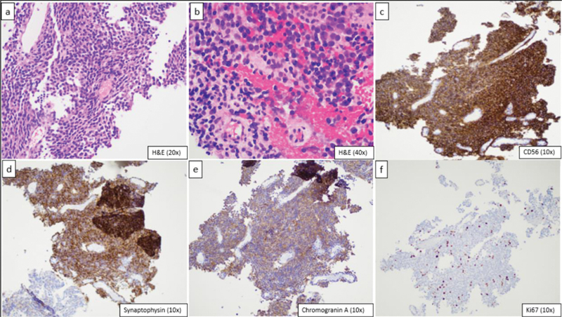
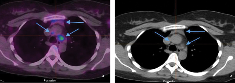
Ectopic pituitary tumors are neoplasms with no connection to the pituitary gland and are commonly deposited in other areas of the anterior skull base. A 32-year-old woman presented with a 3-month history of right-sided facial weakness, sensorineural hearing loss, diplopia, and severe headaches. Physical examination revealed a mid-dilated sluggishly reactive right pupil with slight limitation in all gazes, as well as right-sided orbicularis weakness, lagophthalmos, and decreased facial sensation. A magnetic resonance imaging (MRI) of the head without contrast revealed a 3.7 × 1.8 × 2.6 cm mildly enhancing mass in the right internal acoustic meatus and along the petrous ridge. The case was brought before the institution's tumor board, where concern for higher grade pathology, such as hemangiopericytoma, was discussed. Per patient preference, surgical biopsy of the tumor was performed. Immunohistochemical staining revealed a World Health Organization (WHO) grade II neuroendocrine tumor, with cells staining positive for synaptophysin, chromogranin, and CD56, with a K i -67 index of 8%. In addition to the ectopic location, this pituitary tumor was noted to be aggressive in nature based on its high K i -67 index. Surgical excision and radiologic therapy of tumors involving the CPA are appropriate treatments in most cases.


 求助内容:
求助内容: 应助结果提醒方式:


