Xinrui Huang, Iratxe Torre, Michele Chiappi, Zhan Yin, Anupama Vydyanath, Shuangyi Cao, Oliver Raschdorf, Morgan Beeby, Bonnie Quigley, Pieter P de Tombe, Jun Liu, Edward P Morris, Pradeep K Luther
{"title":"Cryo-electron tomography of intact cardiac muscle reveals myosin binding protein-C linking myosin and actin filaments.","authors":"Xinrui Huang, Iratxe Torre, Michele Chiappi, Zhan Yin, Anupama Vydyanath, Shuangyi Cao, Oliver Raschdorf, Morgan Beeby, Bonnie Quigley, Pieter P de Tombe, Jun Liu, Edward P Morris, Pradeep K Luther","doi":"10.1007/s10974-023-09647-3","DOIUrl":null,"url":null,"abstract":"<p><p>Myosin binding protein C (MyBP-C) is an accessory protein of the thick filament in vertebrate cardiac muscle arranged over 9 stripes of intervals of 430 Å in each half of the A-band in the region called the C-zone. Mutations in cardiac MyBP-C are a leading cause of hypertrophic cardiomyopathy the mechanism of which is unknown. It is a rod-shaped protein composed of 10 or 11 immunoglobulin- or fibronectin-like domains labelled C0 to C10 which binds to the thick filament via its C-terminal region. MyBP-C regulates contraction in a phosphorylation dependent fashion that may be through binding of its N-terminal domains with myosin or actin. Understanding the 3D organisation of MyBP-C in the sarcomere environment may provide new light on its function. We report here the fine structure of MyBP-C in relaxed rat cardiac muscle by cryo-electron tomography and subtomogram averaging of refrozen Tokuyasu cryosections. We find that on average MyBP-C connects via its distal end to actin across a disc perpendicular to the thick filament. The path of MyBP-C suggests that the central domains may interact with myosin heads. Surprisingly MyBP-C at Stripe 4 is different; it has weaker density than the other stripes which could result from a mainly axial or wavy path. Given that the same feature at Stripe 4 can also be found in several mammalian cardiac muscles and in some skeletal muscles, our finding may have broader implication and significance. In the D-zone, we show the first demonstration of myosin crowns arranged on a uniform 143 Å repeat.</p>","PeriodicalId":16422,"journal":{"name":"Journal of Muscle Research and Cell Motility","volume":" ","pages":"165-178"},"PeriodicalIF":1.7000,"publicationDate":"2023-09-01","publicationTypes":"Journal Article","fieldsOfStudy":null,"isOpenAccess":false,"openAccessPdf":"https://www.ncbi.nlm.nih.gov/pmc/articles/PMC10542292/pdf/","citationCount":"0","resultStr":null,"platform":"Semanticscholar","paperid":null,"PeriodicalName":"Journal of Muscle Research and Cell Motility","FirstCategoryId":"99","ListUrlMain":"https://doi.org/10.1007/s10974-023-09647-3","RegionNum":3,"RegionCategory":"生物学","ArticlePicture":[],"TitleCN":null,"AbstractTextCN":null,"PMCID":null,"EPubDate":"2023/4/28 0:00:00","PubModel":"Epub","JCR":"Q4","JCRName":"CELL BIOLOGY","Score":null,"Total":0}
引用次数: 0
Abstract
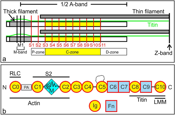
Myosin binding protein C (MyBP-C) is an accessory protein of the thick filament in vertebrate cardiac muscle arranged over 9 stripes of intervals of 430 Å in each half of the A-band in the region called the C-zone. Mutations in cardiac MyBP-C are a leading cause of hypertrophic cardiomyopathy the mechanism of which is unknown. It is a rod-shaped protein composed of 10 or 11 immunoglobulin- or fibronectin-like domains labelled C0 to C10 which binds to the thick filament via its C-terminal region. MyBP-C regulates contraction in a phosphorylation dependent fashion that may be through binding of its N-terminal domains with myosin or actin. Understanding the 3D organisation of MyBP-C in the sarcomere environment may provide new light on its function. We report here the fine structure of MyBP-C in relaxed rat cardiac muscle by cryo-electron tomography and subtomogram averaging of refrozen Tokuyasu cryosections. We find that on average MyBP-C connects via its distal end to actin across a disc perpendicular to the thick filament. The path of MyBP-C suggests that the central domains may interact with myosin heads. Surprisingly MyBP-C at Stripe 4 is different; it has weaker density than the other stripes which could result from a mainly axial or wavy path. Given that the same feature at Stripe 4 can also be found in several mammalian cardiac muscles and in some skeletal muscles, our finding may have broader implication and significance. In the D-zone, we show the first demonstration of myosin crowns arranged on a uniform 143 Å repeat.
期刊介绍:
The Journal of Muscle Research and Cell Motility has as its main aim the publication of original research which bears on either the excitation and contraction of muscle, the analysis of any one of the processes involved therein, the processes underlying contractility and motility of animal and plant cells, the toxicology and pharmacology related to contractility, or the formation, dynamics and turnover of contractile structures in muscle and non-muscle cells. Studies describing the impact of pathogenic mutations in genes encoding components of contractile structures in humans or animals are welcome, provided they offer mechanistic insight into the disease process or the underlying gene function. The policy of the Journal is to encourage any form of novel practical study whatever its specialist interest, as long as it falls within this broad field. Theoretical essays are welcome provided that they are concise and suggest practical ways in which they may be tested. Manuscripts reporting new mutations in known disease genes without validation and mechanistic insight will not be considered. It is the policy of the journal that cells lines, hybridomas and DNA clones should be made available by the developers to any qualified investigator. Submission of a manuscript for publication constitutes an agreement of the authors to abide by this principle.
 求助内容:
求助内容: 应助结果提醒方式:


