{"title":"Virtual biopsy in abdominal pathology: where do we stand?","authors":"Arianna Defeudis, Jovana Panic, Giulia Nicoletti, Simone Mazzetti, Valentina Giannini, Daniele Regge","doi":"10.1259/bjro.20220055","DOIUrl":null,"url":null,"abstract":"<p><p>In recent years, researchers have explored new ways to obtain information from pathological tissues, also exploring non-invasive techniques, such as virtual biopsy (VB). VB can be defined as a test that provides promising outcomes compared to traditional biopsy by extracting quantitative information from radiological images not accessible through traditional visual inspection. Data are processed in such a way that they can be correlated with the patient's phenotypic expression, or with molecular patterns and mutations, creating a bridge between traditional radiology, pathology, genomics, and artificial intelligence (AI). Radiomics is the backbone of VB, since it allows the extraction and selection of features from radiological images, feeding them into AI models in order to derive lesions' pathological characteristics and molecular status. Presently, the output of VB provides only a gross approximation of the findings of tissue biopsy. However, in the future, with the improvement of imaging resolution and processing techniques, VB could partially substitute the classical surgical or percutaneous biopsy, with the advantage of being non-invasive, comprehensive, accounting for lesion heterogeneity, and low cost. In this review, we investigate the concept of VB in abdominal pathology, focusing on its pipeline development and potential benefits.</p>","PeriodicalId":72419,"journal":{"name":"BJR open","volume":"5 1","pages":"20220055"},"PeriodicalIF":2.1000,"publicationDate":"2023-01-01","publicationTypes":"Journal Article","fieldsOfStudy":null,"isOpenAccess":false,"openAccessPdf":"https://www.ncbi.nlm.nih.gov/pmc/articles/PMC10077420/pdf/","citationCount":"0","resultStr":null,"platform":"Semanticscholar","paperid":null,"PeriodicalName":"BJR open","FirstCategoryId":"1085","ListUrlMain":"https://doi.org/10.1259/bjro.20220055","RegionNum":0,"RegionCategory":null,"ArticlePicture":[],"TitleCN":null,"AbstractTextCN":null,"PMCID":null,"EPubDate":"","PubModel":"","JCR":"","JCRName":"","Score":null,"Total":0}
引用次数: 0
Abstract
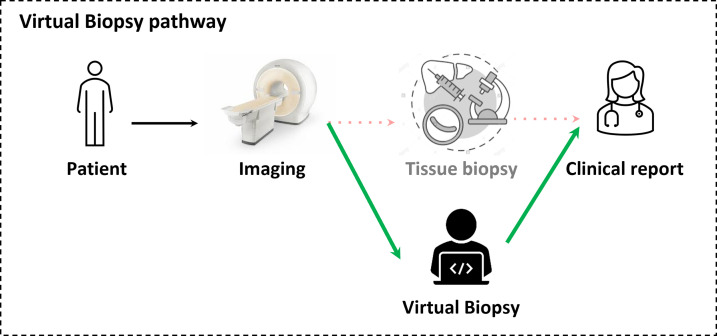
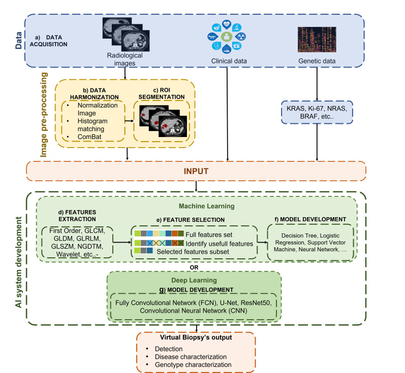
In recent years, researchers have explored new ways to obtain information from pathological tissues, also exploring non-invasive techniques, such as virtual biopsy (VB). VB can be defined as a test that provides promising outcomes compared to traditional biopsy by extracting quantitative information from radiological images not accessible through traditional visual inspection. Data are processed in such a way that they can be correlated with the patient's phenotypic expression, or with molecular patterns and mutations, creating a bridge between traditional radiology, pathology, genomics, and artificial intelligence (AI). Radiomics is the backbone of VB, since it allows the extraction and selection of features from radiological images, feeding them into AI models in order to derive lesions' pathological characteristics and molecular status. Presently, the output of VB provides only a gross approximation of the findings of tissue biopsy. However, in the future, with the improvement of imaging resolution and processing techniques, VB could partially substitute the classical surgical or percutaneous biopsy, with the advantage of being non-invasive, comprehensive, accounting for lesion heterogeneity, and low cost. In this review, we investigate the concept of VB in abdominal pathology, focusing on its pipeline development and potential benefits.

 求助内容:
求助内容: 应助结果提醒方式:


