Soluble PD‑L1 reflects cachexia status in patients with gastric cancer and is an independent prognostic marker for relapse‑free survival after radical surgery.
{"title":"Soluble PD‑L1 reflects cachexia status in patients with gastric cancer and is an independent prognostic marker for relapse‑free survival after radical surgery.","authors":"Yasunori Matsumoto, Takuma Sasaki, Masayuki Kano, Tadashi Shiraishi, Hiroshi Suito, Kentaro Murakami, Takeshi Toyozumi, Ryota Otsuka, Kazuya Kinoshita, Shinichiro Iida, Hiroki Morishita, Yuri Nishioka, Koichi Hayano, Yoshihiro Kurata, Hideki Hayashi, Hisahiro Matsubara","doi":"10.3892/mco.2023.2635","DOIUrl":null,"url":null,"abstract":"<p><p>Soluble programmed death-ligand 1 (sPD-L1) levels can be used as a biomarker for gastric cancer (GC). However, comprehensive information regarding the sPD-L1 expression profiles and their association with cachexia in GC is lacking. Therefore, the present study evaluated the association between clinicopathological findings and sPD-L1 levels in patients with GC. Serum samples were collected from patients with GC during their first visit to Department of Esophageal-Gastro-Intestinal Surgery, Chiba University Hospital, Chiba, Japan (January 2012-December 2017; n=173), and sPD-L1 levels were measured using an enzyme-linked immunosorbent assay. Survival rates among 116 patients, excluding cases with preoperative chemotherapy or no radical procedures, were analyzed. sPD-L1 levels were associated with factors such as neutrophil-to-lymphocyte ratio, hemoglobin (Hb) and albumin (Alb) levels, total cholesterol and C-reactive protein (CRP) levels, and related to inflammation and nutrition in patients. Notably, the higher the number of applicable indicators related to cachexia (Hb <12 g/dl, Alb <3.2 g/dl, CRP >0.5 mg/dl and low body mass index) was, the higher the sPD-L1 value was. However, the pathological stage did not significantly differ between the groups. Clinicopathologically, there was no association with tumor depth, lymph node metastasis or vascular invasion; however, patients with the intestinal type had significantly higher sPD-L1 levels than patients with the diffuse type (P=0.032; Wilcoxon test). The overall survival did not significantly differ between the groups with low and high sPD-L1 levels; however, among patients who received radical treatment, the relapse-free survival was significantly worse in the high-sPD-L1-level group than in the low-sPD-L1-level group (P=0.025; log-rank test). Multivariate Cox regression analysis revealed that a high sPD-L1 concentration was a sign of poor prognosis, independent of pathological stage and cancer antigen CA19-9 (P=0.0029). Therefore, the present findings suggest that sPD-L1 can reflect cachexia status in patients with GC and may serve as a prognostic marker for relapse-free survival after radical GC surgery.</p>","PeriodicalId":18737,"journal":{"name":"Molecular and clinical oncology","volume":"18 5","pages":"39"},"PeriodicalIF":1.4000,"publicationDate":"2023-05-01","publicationTypes":"Journal Article","fieldsOfStudy":null,"isOpenAccess":false,"openAccessPdf":"https://ftp.ncbi.nlm.nih.gov/pub/pmc/oa_pdf/70/2a/mco-18-05-02635.PMC10074020.pdf","citationCount":"1","resultStr":null,"platform":"Semanticscholar","paperid":null,"PeriodicalName":"Molecular and clinical oncology","FirstCategoryId":"1085","ListUrlMain":"https://doi.org/10.3892/mco.2023.2635","RegionNum":0,"RegionCategory":null,"ArticlePicture":[],"TitleCN":null,"AbstractTextCN":null,"PMCID":null,"EPubDate":"","PubModel":"","JCR":"Q4","JCRName":"ONCOLOGY","Score":null,"Total":0}
引用次数: 1
Abstract
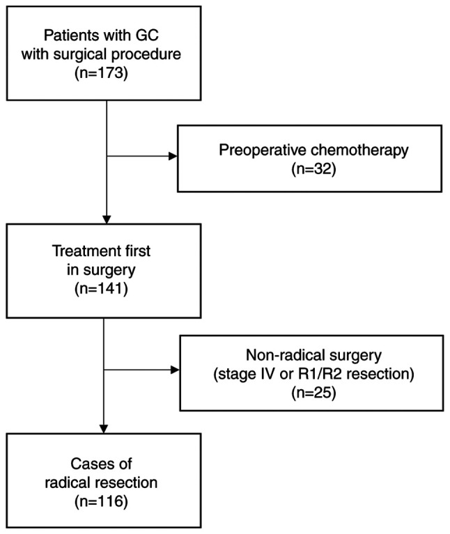
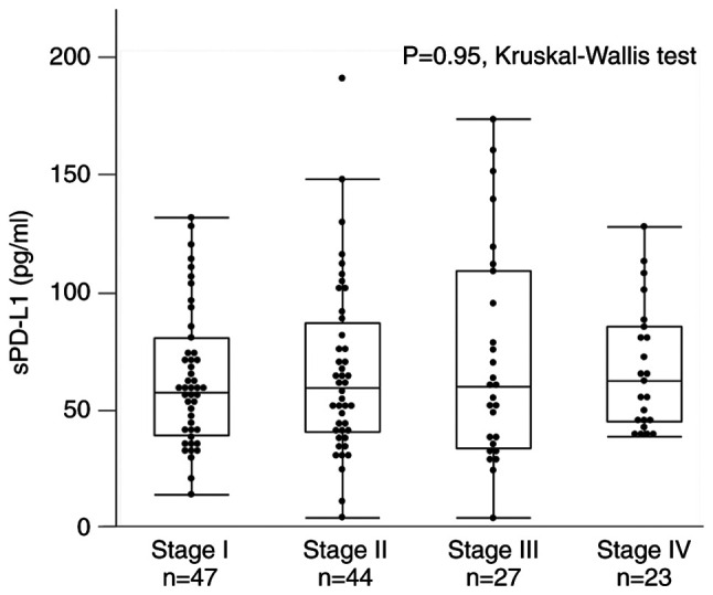
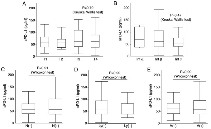
Soluble programmed death-ligand 1 (sPD-L1) levels can be used as a biomarker for gastric cancer (GC). However, comprehensive information regarding the sPD-L1 expression profiles and their association with cachexia in GC is lacking. Therefore, the present study evaluated the association between clinicopathological findings and sPD-L1 levels in patients with GC. Serum samples were collected from patients with GC during their first visit to Department of Esophageal-Gastro-Intestinal Surgery, Chiba University Hospital, Chiba, Japan (January 2012-December 2017; n=173), and sPD-L1 levels were measured using an enzyme-linked immunosorbent assay. Survival rates among 116 patients, excluding cases with preoperative chemotherapy or no radical procedures, were analyzed. sPD-L1 levels were associated with factors such as neutrophil-to-lymphocyte ratio, hemoglobin (Hb) and albumin (Alb) levels, total cholesterol and C-reactive protein (CRP) levels, and related to inflammation and nutrition in patients. Notably, the higher the number of applicable indicators related to cachexia (Hb <12 g/dl, Alb <3.2 g/dl, CRP >0.5 mg/dl and low body mass index) was, the higher the sPD-L1 value was. However, the pathological stage did not significantly differ between the groups. Clinicopathologically, there was no association with tumor depth, lymph node metastasis or vascular invasion; however, patients with the intestinal type had significantly higher sPD-L1 levels than patients with the diffuse type (P=0.032; Wilcoxon test). The overall survival did not significantly differ between the groups with low and high sPD-L1 levels; however, among patients who received radical treatment, the relapse-free survival was significantly worse in the high-sPD-L1-level group than in the low-sPD-L1-level group (P=0.025; log-rank test). Multivariate Cox regression analysis revealed that a high sPD-L1 concentration was a sign of poor prognosis, independent of pathological stage and cancer antigen CA19-9 (P=0.0029). Therefore, the present findings suggest that sPD-L1 can reflect cachexia status in patients with GC and may serve as a prognostic marker for relapse-free survival after radical GC surgery.

 求助内容:
求助内容: 应助结果提醒方式:


