Retinal pathological features and proteome signatures of Alzheimer’s disease

IF 9.3 1区 医学 Q1 CLINICAL NEUROLOGY
Yosef Koronyo, Altan Rentsendorj, Nazanin Mirzaei, Giovanna C. Regis, Julia Sheyn, Haoshen Shi, Ernesto Barron, Galen Cook-Wiens, Anthony R. Rodriguez, Rodrigo Medeiros, Joao A. Paulo, Veer B. Gupta, Andrei A. Kramerov, Alexander V. Ljubimov, Jennifer E. Van Eyk, Stuart L. Graham, Vivek K. Gupta, John M. Ringman, David R. Hinton, Carol A. Miller, Keith L. Black, Antonino Cattaneo, Giovanni Meli, Mehdi Mirzaei, Dieu-Trang Fuchs, Maya Koronyo-Hamaoui
{"title":"Retinal pathological features and proteome signatures of Alzheimer’s disease","authors":"Yosef Koronyo,&nbsp;Altan Rentsendorj,&nbsp;Nazanin Mirzaei,&nbsp;Giovanna C. Regis,&nbsp;Julia Sheyn,&nbsp;Haoshen Shi,&nbsp;Ernesto Barron,&nbsp;Galen Cook-Wiens,&nbsp;Anthony R. Rodriguez,&nbsp;Rodrigo Medeiros,&nbsp;Joao A. Paulo,&nbsp;Veer B. Gupta,&nbsp;Andrei A. Kramerov,&nbsp;Alexander V. Ljubimov,&nbsp;Jennifer E. Van Eyk,&nbsp;Stuart L. Graham,&nbsp;Vivek K. Gupta,&nbsp;John M. Ringman,&nbsp;David R. Hinton,&nbsp;Carol A. Miller,&nbsp;Keith L. Black,&nbsp;Antonino Cattaneo,&nbsp;Giovanni Meli,&nbsp;Mehdi Mirzaei,&nbsp;Dieu-Trang Fuchs,&nbsp;Maya Koronyo-Hamaoui","doi":"10.1007/s00401-023-02548-2","DOIUrl":null,"url":null,"abstract":"<div><p>Alzheimer’s disease (AD) pathologies were discovered in the accessible neurosensory retina. However, their exact nature and topographical distribution, particularly in the early stages of functional impairment, and how they relate to disease progression in the brain remain largely unknown. To better understand the pathological features of AD in the retina, we conducted an extensive histopathological and biochemical investigation of postmortem retina and brain tissues from 86 human donors. Quantitative examination of superior and inferior temporal retinas from mild cognitive impairment (MCI) and AD patients compared to those with normal cognition (NC) revealed significant increases in amyloid β-protein (Aβ<sub>42</sub>) forms and novel intraneuronal Aβ oligomers (AβOi), which were closely associated with exacerbated retinal macrogliosis, microgliosis, and tissue atrophy. These pathologies were unevenly distributed across retinal layers and geometrical areas, with the inner layers and peripheral subregions exhibiting most pronounced accumulations in the MCI and AD versus NC retinas. While microgliosis was increased in the retina of these patients, the proportion of microglial cells engaging in Aβ uptake was reduced. Female AD patients exhibited higher levels of retinal microgliosis than males. Notably, retinal Aβ<sub>42</sub>, S100 calcium-binding protein B<sup>+</sup> macrogliosis, and atrophy correlated with severity of brain Aβ pathology, tauopathy, and atrophy, and most retinal pathologies reflected Braak staging. All retinal biomarkers correlated with the cognitive scores, with retinal Aβ<sub>42</sub>, far-peripheral AβOi and microgliosis displaying the strongest correlations. Proteomic analysis of AD retinas revealed activation of specific inflammatory and neurodegenerative processes and inhibition of oxidative phosphorylation/mitochondrial, and photoreceptor-related pathways. This study identifies and maps retinopathy in MCI and AD patients, demonstrating the quantitative relationship with brain pathology and cognition, and may lead to reliable retinal biomarkers for noninvasive retinal screening and monitoring of AD.</p></div>","PeriodicalId":7012,"journal":{"name":"Acta Neuropathologica","volume":"145 4","pages":"409 - 438"},"PeriodicalIF":9.3000,"publicationDate":"2023-02-11","publicationTypes":"Journal Article","fieldsOfStudy":null,"isOpenAccess":false,"openAccessPdf":"https://link.springer.com/content/pdf/10.1007/s00401-023-02548-2.pdf","citationCount":"11","resultStr":null,"platform":"Semanticscholar","paperid":null,"PeriodicalName":"Acta Neuropathologica","FirstCategoryId":"3","ListUrlMain":"https://link.springer.com/article/10.1007/s00401-023-02548-2","RegionNum":1,"RegionCategory":"医学","ArticlePicture":[],"TitleCN":null,"AbstractTextCN":null,"PMCID":null,"EPubDate":"","PubModel":"","JCR":"Q1","JCRName":"CLINICAL NEUROLOGY","Score":null,"Total":0}
引用次数: 11

Abstract

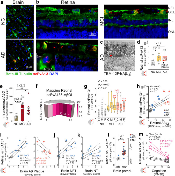
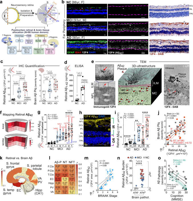
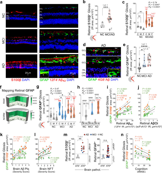
Alzheimer’s disease (AD) pathologies were discovered in the accessible neurosensory retina. However, their exact nature and topographical distribution, particularly in the early stages of functional impairment, and how they relate to disease progression in the brain remain largely unknown. To better understand the pathological features of AD in the retina, we conducted an extensive histopathological and biochemical investigation of postmortem retina and brain tissues from 86 human donors. Quantitative examination of superior and inferior temporal retinas from mild cognitive impairment (MCI) and AD patients compared to those with normal cognition (NC) revealed significant increases in amyloid β-protein (Aβ42) forms and novel intraneuronal Aβ oligomers (AβOi), which were closely associated with exacerbated retinal macrogliosis, microgliosis, and tissue atrophy. These pathologies were unevenly distributed across retinal layers and geometrical areas, with the inner layers and peripheral subregions exhibiting most pronounced accumulations in the MCI and AD versus NC retinas. While microgliosis was increased in the retina of these patients, the proportion of microglial cells engaging in Aβ uptake was reduced. Female AD patients exhibited higher levels of retinal microgliosis than males. Notably, retinal Aβ42, S100 calcium-binding protein B+ macrogliosis, and atrophy correlated with severity of brain Aβ pathology, tauopathy, and atrophy, and most retinal pathologies reflected Braak staging. All retinal biomarkers correlated with the cognitive scores, with retinal Aβ42, far-peripheral AβOi and microgliosis displaying the strongest correlations. Proteomic analysis of AD retinas revealed activation of specific inflammatory and neurodegenerative processes and inhibition of oxidative phosphorylation/mitochondrial, and photoreceptor-related pathways. This study identifies and maps retinopathy in MCI and AD patients, demonstrating the quantitative relationship with brain pathology and cognition, and may lead to reliable retinal biomarkers for noninvasive retinal screening and monitoring of AD.
Abstract Image

Abstract Image

Abstract Image

阿尔茨海默病的视网膜病理特征和蛋白质组特征
阿尔茨海默病(AD)病变是在可接近的神经感觉视网膜中发现的。然而,它们的确切性质和地形分布,特别是在功能损伤的早期阶段,以及它们与大脑疾病进展的关系,在很大程度上仍然未知。为了更好地了解AD在视网膜中的病理特征,我们对86名人类供体的死后视网膜和脑组织进行了广泛的组织病理学和生化研究。与认知正常(NC)患者相比,轻度认知障碍(MCI)和AD患者颞叶上下视网膜的定量检查显示,淀粉样蛋白(Aβ42)形式和新型神经内Aβ低聚物(AβOi)显著增加,这与视网膜大胶质细胞增生、小胶质细胞增生和组织萎缩的加剧密切相关。这些病变在视网膜层和几何区域中分布不均,内层和外围亚区在MCI和AD视网膜中表现出最明显的积聚。当这些患者的视网膜中小胶质细胞增多时,参与Aβ摄取的小胶质细胞比例降低。女性AD患者的视网膜小胶质细胞增生水平高于男性。值得注意的是,视网膜Aβ42、S100钙结合蛋白B+大胶质细胞增生和萎缩与大脑Aβ病理、tau病和萎缩的严重程度相关,大多数视网膜病理反映了Braak分期。所有视网膜生物标志物都与认知评分相关,其中视网膜Aβ42、远外周AβOi和小胶质细胞增生显示出最强的相关性。AD视网膜的蛋白质组学分析揭示了特定炎症和神经退行性过程的激活以及氧化磷酸化/线粒体和光感受器相关途径的抑制。这项研究识别并绘制了MCI和AD患者的视网膜病变,证明了其与大脑病理和认知的定量关系,并可能为AD的非侵入性视网膜筛查和监测提供可靠的视网膜生物标志物。
本文章由计算机程序翻译,如有差异,请以英文原文为准。
求助全文
约1分钟内获得全文 求助全文
来源期刊
Acta Neuropathologica
Acta Neuropathologica 医学-病理学
CiteScore
23.70
自引率
3.90%
发文量
118
审稿时长
4-8 weeks
期刊介绍: Acta Neuropathologica publishes top-quality papers on the pathology of neurological diseases and experimental studies on molecular and cellular mechanisms using in vitro and in vivo models, ideally validated by analysis of human tissues. The journal accepts Original Papers, Review Articles, Case Reports, and Scientific Correspondence (Letters). Manuscripts must adhere to ethical standards, including review by appropriate ethics committees for human studies and compliance with principles of laboratory animal care for animal experiments. Failure to comply may result in rejection of the manuscript, and authors are responsible for ensuring accuracy and adherence to these requirements.
×
引用
GB/T 7714-2015
复制
MLA
复制
APA
复制
导出至
BibTeX EndNote RefMan NoteFirst NoteExpress
×
提示
您的信息不完整,为了账户安全,请先补充。
现在去补充
×
提示
您因"违规操作"
具体请查看互助需知
我知道了
×
提示
确定
请完成安全验证×
copy
已复制链接
快去分享给好友吧!
我知道了
右上角分享
点击右上角分享
0
联系我们:info@booksci.cn Book学术提供免费学术资源搜索服务,方便国内外学者检索中英文文献。致力于提供最便捷和优质的服务体验。 Copyright © 2023 布克学术 All rights reserved.
京ICP备2023020795号-1
ghs 京公网安备 11010802042870号
Book学术文献互助
Book学术文献互助群
群 号:481959085
Book学术官方微信