Ueli Braun, Christian Gerspach, Claudia Volz, Muriel Boesiger, Monika Hilbe, Karl Nuss
{"title":"A retrospective review of small intestinal intussusception in 126 cattle in Switzerland.","authors":"Ueli Braun, Christian Gerspach, Claudia Volz, Muriel Boesiger, Monika Hilbe, Karl Nuss","doi":"10.1002/vro2.58","DOIUrl":null,"url":null,"abstract":"<p><strong>Background: </strong>Intussusception is a form of ileus of the intestines in which an oral intestinal segment slides into the adjacent aboral intestinal segment, causing obstruction of the bowel.</p><p><strong>Methods: </strong>We analysed the medical records of 126 cattle with intussusception of the small intestine.</p><p><strong>Results: </strong>Demeanour and appetite were abnormal in 123 cattle. Non-specific signs of pain occurred in 26.2%, signs of visceral pain in 46.8% and signs of parietal pain in 56.4%. Intestinal motility was decreased or absent in 93.7% of the cattle. The most common findings of transrectal palpation were rumen dilation (37.3%) and dilated small intestines (24.6%). In 96% of the cattle, the rectum was empty or contained little faeces. The principal laboratory findings were hypokalaemia (89.6%), hypocalcaemia (76.5%), base excess (72.9%), hypochloraemia (71.8%), azotaemia (62.1%) and haemoconcentration (61.1%). The main ultrasonographic findings were reduced or absent intestinal motility (98.2%) and dilated small intestines (96.0%). A diagnosis of ileus was made in 87.8% and a diagnosis of ileus attributable to intussusception was made in another 9.8%. Right-flank laparotomy was carried out in 114 cattle. Fifty-six (44.4%) cows were discharged.</p><p><strong>Conclusions: </strong>Clinical findings of intussusception in cattle are often non-specific. Ultrasonography may be required to diagnose ileus.</p>","PeriodicalId":23565,"journal":{"name":"Veterinary Record Open","volume":"10 1","pages":"e58"},"PeriodicalIF":1.1000,"publicationDate":"2023-03-28","publicationTypes":"Journal Article","fieldsOfStudy":null,"isOpenAccess":false,"openAccessPdf":"https://www.ncbi.nlm.nih.gov/pmc/articles/PMC10049975/pdf/","citationCount":"0","resultStr":null,"platform":"Semanticscholar","paperid":null,"PeriodicalName":"Veterinary Record Open","FirstCategoryId":"1085","ListUrlMain":"https://doi.org/10.1002/vro2.58","RegionNum":0,"RegionCategory":null,"ArticlePicture":[],"TitleCN":null,"AbstractTextCN":null,"PMCID":null,"EPubDate":"2023/6/1 0:00:00","PubModel":"eCollection","JCR":"Q2","JCRName":"VETERINARY SCIENCES","Score":null,"Total":0}
引用次数: 0
Abstract
Background: Intussusception is a form of ileus of the intestines in which an oral intestinal segment slides into the adjacent aboral intestinal segment, causing obstruction of the bowel.
Methods: We analysed the medical records of 126 cattle with intussusception of the small intestine.
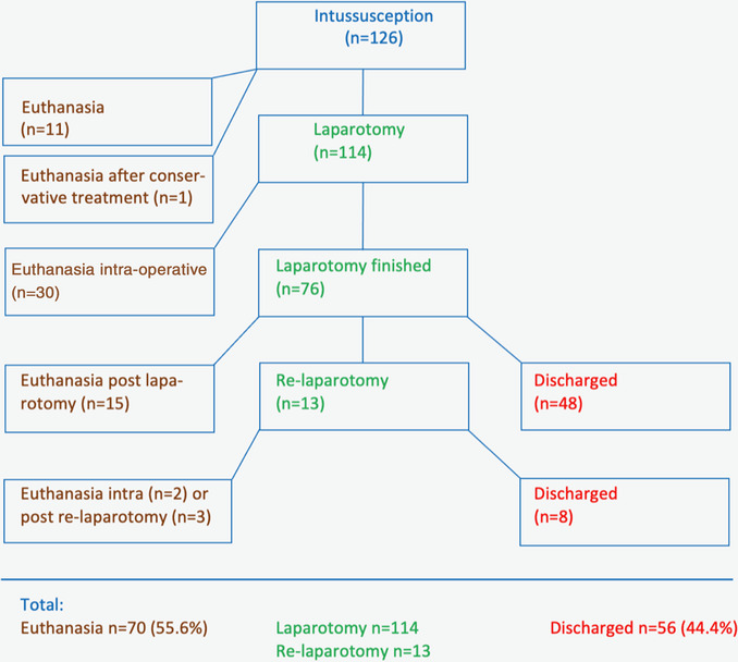
Results: Demeanour and appetite were abnormal in 123 cattle. Non-specific signs of pain occurred in 26.2%, signs of visceral pain in 46.8% and signs of parietal pain in 56.4%. Intestinal motility was decreased or absent in 93.7% of the cattle. The most common findings of transrectal palpation were rumen dilation (37.3%) and dilated small intestines (24.6%). In 96% of the cattle, the rectum was empty or contained little faeces. The principal laboratory findings were hypokalaemia (89.6%), hypocalcaemia (76.5%), base excess (72.9%), hypochloraemia (71.8%), azotaemia (62.1%) and haemoconcentration (61.1%). The main ultrasonographic findings were reduced or absent intestinal motility (98.2%) and dilated small intestines (96.0%). A diagnosis of ileus was made in 87.8% and a diagnosis of ileus attributable to intussusception was made in another 9.8%. Right-flank laparotomy was carried out in 114 cattle. Fifty-six (44.4%) cows were discharged.
Conclusions: Clinical findings of intussusception in cattle are often non-specific. Ultrasonography may be required to diagnose ileus.
期刊介绍:
Veterinary Record Open is a journal dedicated to publishing specialist veterinary research across a range of topic areas including those of a more niche and specialist nature to that considered in the weekly Vet Record. Research from all disciplines of veterinary interest will be considered. It is an Open Access journal of the British Veterinary Association.

 求助内容:
求助内容: 应助结果提醒方式:


