Breath-hold High-resolution T1-weighted Gradient Echo Liver MR Imaging with Compressed Sensing Obtained during the Gadoxetic Acid-enhanced Hepatobiliary Phase: Image Quality and Lesion Visibility Compared with a Standard T1-weighted Sequence.
IF 3.2 3区 医学Q2 RADIOLOGY, NUCLEAR MEDICINE & MEDICAL IMAGING
{"title":"Breath-hold High-resolution T1-weighted Gradient Echo Liver MR Imaging with Compressed Sensing Obtained during the Gadoxetic Acid-enhanced Hepatobiliary Phase: Image Quality and Lesion Visibility Compared with a Standard T1-weighted Sequence.","authors":"Kenichiro Ihara, Hideko Onoda, Masahiro Tanabe, Etsushi Iida, Takaaki Ueda, Taiga Kobayashi, Mayumi Higashi, Marcel Dominik Nickel, Hiroshi Imai, Katsuyoshi Ito","doi":"10.2463/mrms.mp.2022-0137","DOIUrl":null,"url":null,"abstract":"<p><strong>Purpose: </strong>To evaluate the feasibility of breath-hold (BH) high-resolution (HR) T1-weighted gradient echo hepatobiliary phase (HBP) imaging using compressed sensing (CS) in gadoxetic acid-enhanced liver MRI in comparison with standard HBP imaging using parallel imaging (PI).</p><p><strong>Methods: </strong>The study included 122 patients with liver tumors with hypointensity in the HBP who underwent both HR HBP imaging with CS and standard HBP imaging with PI. Two radiologists evaluated the liver edge sharpness, hepatic vessel conspicuity, bile duct conspicuity, image noise, and overall image quality, as well as the lesion conspicuity on HR and standard HBP imaging and the contrast-enhanced (CE) MR cholangiography (MRC) image quality reconstructed from HBP images. As a quantitative analysis, the SNR of the liver and the liver to lesion signal intensity ratio (LLSIR) were also determined.</p><p><strong>Results: </strong>The liver edge sharpness, hepatic vessel conspicuity, bile duct conspicuity, and overall image quality as well as the lesion conspicuity and the LLSIR on HR HBP imaging with CS were significantly higher than those on standard HBP imaging (all of P < 0.001). The image quality of CE-MRC reconstructed from HR HBP imaging with CS was also significantly higher than that from standard HBP imaging (P < 0.001). Conversely, the SNR of liver in standard HBP was significantly higher than that in HR HBP with CS (P < 0.001).</p><p><strong>Conclusion: </strong>BH HR HBP imaging with CS provided an improved overall image quality, lesion conspicuity, and CE-MRC visualization when compared with standard HBP imaging without extending the acquisition time.</p>","PeriodicalId":18119,"journal":{"name":"Magnetic Resonance in Medical Sciences","volume":" ","pages":"146-152"},"PeriodicalIF":3.2000,"publicationDate":"2024-04-01","publicationTypes":"Journal Article","fieldsOfStudy":null,"isOpenAccess":false,"openAccessPdf":"https://www.ncbi.nlm.nih.gov/pmc/articles/PMC11024715/pdf/","citationCount":"0","resultStr":null,"platform":"Semanticscholar","paperid":null,"PeriodicalName":"Magnetic Resonance in Medical Sciences","FirstCategoryId":"3","ListUrlMain":"https://doi.org/10.2463/mrms.mp.2022-0137","RegionNum":3,"RegionCategory":"医学","ArticlePicture":[],"TitleCN":null,"AbstractTextCN":null,"PMCID":null,"EPubDate":"2023/2/4 0:00:00","PubModel":"Epub","JCR":"Q2","JCRName":"RADIOLOGY, NUCLEAR MEDICINE & MEDICAL IMAGING","Score":null,"Total":0}
引用次数: 0
Abstract
Purpose: To evaluate the feasibility of breath-hold (BH) high-resolution (HR) T1-weighted gradient echo hepatobiliary phase (HBP) imaging using compressed sensing (CS) in gadoxetic acid-enhanced liver MRI in comparison with standard HBP imaging using parallel imaging (PI).
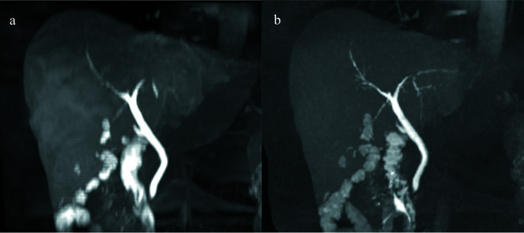
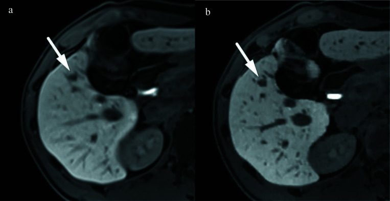
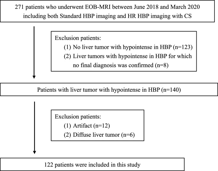
Methods: The study included 122 patients with liver tumors with hypointensity in the HBP who underwent both HR HBP imaging with CS and standard HBP imaging with PI. Two radiologists evaluated the liver edge sharpness, hepatic vessel conspicuity, bile duct conspicuity, image noise, and overall image quality, as well as the lesion conspicuity on HR and standard HBP imaging and the contrast-enhanced (CE) MR cholangiography (MRC) image quality reconstructed from HBP images. As a quantitative analysis, the SNR of the liver and the liver to lesion signal intensity ratio (LLSIR) were also determined.
Results: The liver edge sharpness, hepatic vessel conspicuity, bile duct conspicuity, and overall image quality as well as the lesion conspicuity and the LLSIR on HR HBP imaging with CS were significantly higher than those on standard HBP imaging (all of P < 0.001). The image quality of CE-MRC reconstructed from HR HBP imaging with CS was also significantly higher than that from standard HBP imaging (P < 0.001). Conversely, the SNR of liver in standard HBP was significantly higher than that in HR HBP with CS (P < 0.001).
Conclusion: BH HR HBP imaging with CS provided an improved overall image quality, lesion conspicuity, and CE-MRC visualization when compared with standard HBP imaging without extending the acquisition time.
期刊介绍:
Magnetic Resonance in Medical Sciences (MRMS or Magn
Reson Med Sci) is an international journal pursuing the
publication of original articles contributing to the progress
of magnetic resonance in the field of biomedical sciences
including technical developments and clinical applications.
MRMS is an official journal of the Japanese Society for
Magnetic Resonance in Medicine (JSMRM).
 求助内容:
求助内容: 应助结果提醒方式:


